Cytoplasmic Ana Pattern
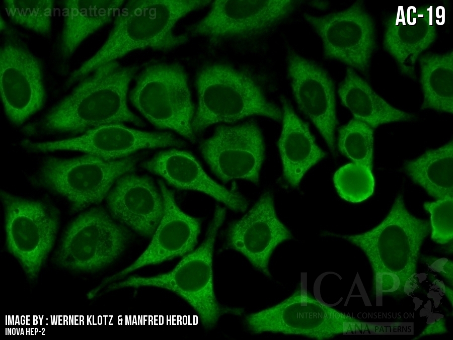
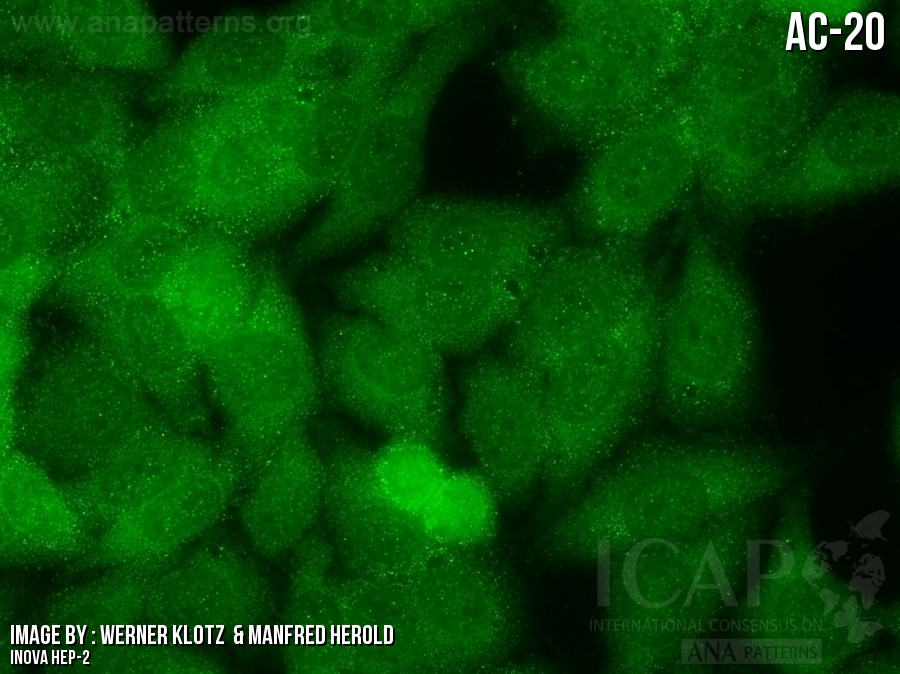
Cytoplasmic Ana Pattern - This pattern is characterized by decorated cytoskeletal fibers, sometimes with small, discontinuous granular deposits. A titer (a measure of how much ana is in the blood) and a pattern (where the ana was detected in the cells). May be associated with hepatitis, hepatitis c, lupus, myositis, or sometimes mean nothing at all. Web welcome to anapatterns.org, the official website for the international consensus on antinuclear antibody (ana) patterns (icap). The dense fine speckled pattern. Typical staining show striated actin cables spanning the long axis of the cells. Ana test results provide patterns that may be suggestive of a. Web antinuclear antibody (ana) testing is useful as an initial screen for autoimmune diseases such as sjögren syndrome, systemic lupus erythematosus, and scleroderma. One pattern that deserves special attention is. Negative (n = 1), nuclear (n = 15), cytoplasmic (n = 9), and mitotic patterns (n = 5) (2, 3, 5, 7, 8). Negative (n = 1), nuclear (n = 15), cytoplasmic (n = 9), and mitotic patterns (n = 5) (2, 3, 5, 7, 8). Web welcome to anapatterns.org, the official website for the international consensus on antinuclear antibody (ana) patterns (icap). Typical staining show striated actin cables spanning the long axis of the cells. May be associated with hepatitis, hepatitis c,. One pattern that deserves special attention is. Web welcome to anapatterns.org, the official website for the international consensus on antinuclear antibody (ana) patterns (icap). Negative (n = 1), nuclear (n = 15), cytoplasmic (n = 9), and mitotic patterns (n = 5) (2, 3, 5, 7, 8). May be associated with hepatitis, hepatitis c, lupus, myositis, or sometimes mean nothing. This pattern is characterized by decorated cytoskeletal fibers, sometimes with small, discontinuous granular deposits. A titer (a measure of how much ana is in the blood) and a pattern (where the ana was detected in the cells). One pattern that deserves special attention is. Typical staining show striated actin cables spanning the long axis of the cells. May be associated. A titer (a measure of how much ana is in the blood) and a pattern (where the ana was detected in the cells). This pattern is characterized by decorated cytoskeletal fibers, sometimes with small, discontinuous granular deposits. Ana test results provide patterns that may be suggestive of a. Typical staining show striated actin cables spanning the long axis of the. Typical staining show striated actin cables spanning the long axis of the cells. A titer (a measure of how much ana is in the blood) and a pattern (where the ana was detected in the cells). Web antinuclear antibody (ana) testing is useful as an initial screen for autoimmune diseases such as sjögren syndrome, systemic lupus erythematosus, and scleroderma. Web. Negative (n = 1), nuclear (n = 15), cytoplasmic (n = 9), and mitotic patterns (n = 5) (2, 3, 5, 7, 8). Web antinuclear antibody (ana) testing is useful as an initial screen for autoimmune diseases such as sjögren syndrome, systemic lupus erythematosus, and scleroderma. The dense fine speckled pattern. Typical staining show striated actin cables spanning the long. One pattern that deserves special attention is. Typical staining show striated actin cables spanning the long axis of the cells. Negative (n = 1), nuclear (n = 15), cytoplasmic (n = 9), and mitotic patterns (n = 5) (2, 3, 5, 7, 8). This pattern is characterized by decorated cytoskeletal fibers, sometimes with small, discontinuous granular deposits. A titer (a. Typical staining show striated actin cables spanning the long axis of the cells. May be associated with hepatitis, hepatitis c, lupus, myositis, or sometimes mean nothing at all. The dense fine speckled pattern. One pattern that deserves special attention is. Negative (n = 1), nuclear (n = 15), cytoplasmic (n = 9), and mitotic patterns (n = 5) (2, 3,. May be associated with hepatitis, hepatitis c, lupus, myositis, or sometimes mean nothing at all. A titer (a measure of how much ana is in the blood) and a pattern (where the ana was detected in the cells). The dense fine speckled pattern. Web antinuclear antibody (ana) testing is useful as an initial screen for autoimmune diseases such as sjögren. Web welcome to anapatterns.org, the official website for the international consensus on antinuclear antibody (ana) patterns (icap). One pattern that deserves special attention is. Web antinuclear antibody (ana) testing is useful as an initial screen for autoimmune diseases such as sjögren syndrome, systemic lupus erythematosus, and scleroderma. Negative (n = 1), nuclear (n = 15), cytoplasmic (n = 9), and. Web antinuclear antibody (ana) testing is useful as an initial screen for autoimmune diseases such as sjögren syndrome, systemic lupus erythematosus, and scleroderma. Web welcome to anapatterns.org, the official website for the international consensus on antinuclear antibody (ana) patterns (icap). Ana test results provide patterns that may be suggestive of a. The dense fine speckled pattern. One pattern that deserves special attention is. Negative (n = 1), nuclear (n = 15), cytoplasmic (n = 9), and mitotic patterns (n = 5) (2, 3, 5, 7, 8). Typical staining show striated actin cables spanning the long axis of the cells. This pattern is characterized by decorated cytoskeletal fibers, sometimes with small, discontinuous granular deposits.
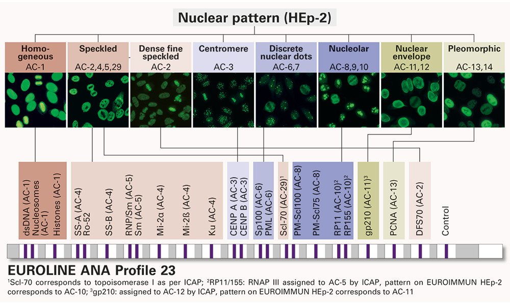
Multiplex determination of ANA and cytoplasmic antibodies according to

ANA Patterns

ANA Patterns

ANA Patterns

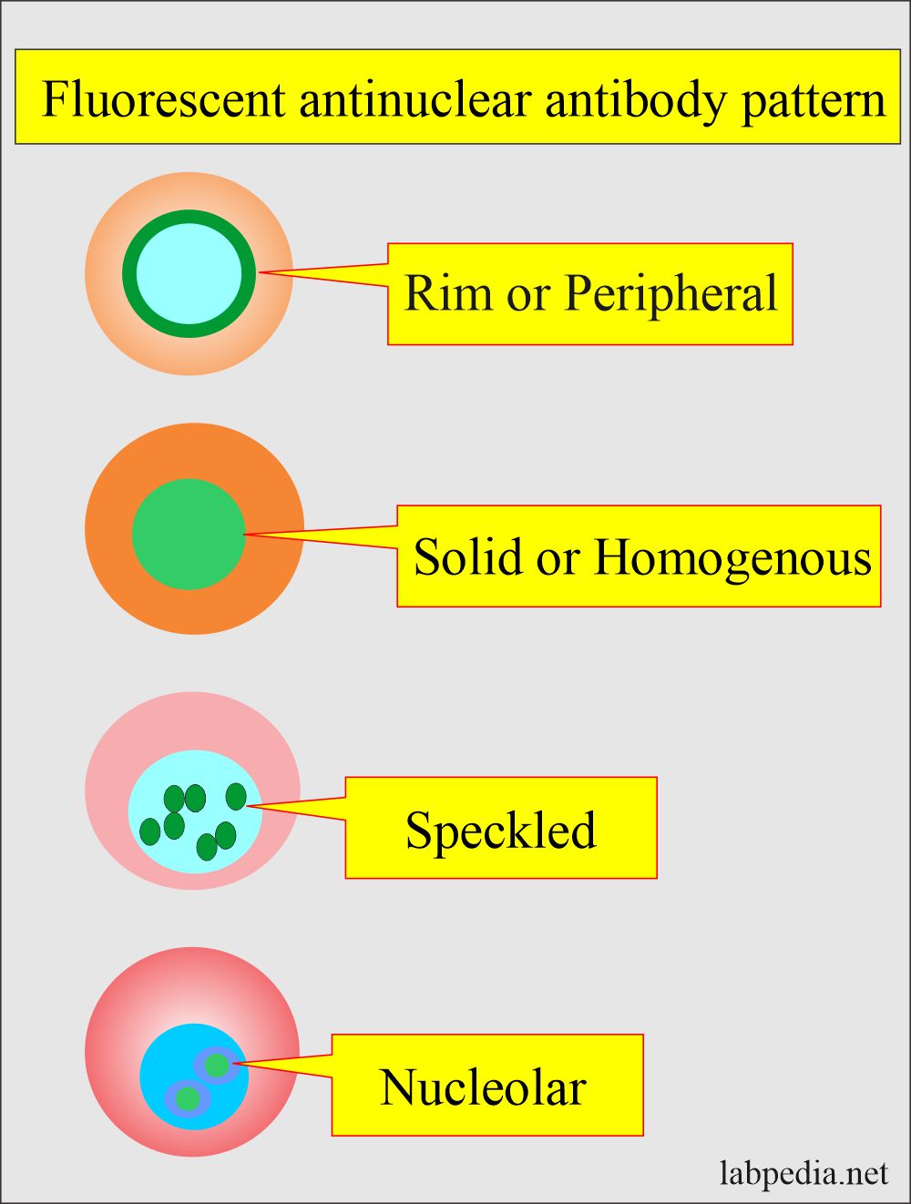
Antinuclear Factor (ANF), Antinuclear Antibody (ANA) and Its

ANA Patterns

ANA Patterns

ANA Patterns

Cytoplasmic Ana Pattern 7thongs

ANA Patterns
A Titer (A Measure Of How Much Ana Is In The Blood) And A Pattern (Where The Ana Was Detected In The Cells).
May Be Associated With Hepatitis, Hepatitis C, Lupus, Myositis, Or Sometimes Mean Nothing At All.
Related Post: