Tissue Drawing
Tissue Drawing - Web at the end of this article, there is the drawing procedure and images of the simple squamous. This is made up of thin, flat and hexagonal cells. Web beginning a&p content, organ systems, directional terms, body sections and cavities, epithelial, connective, muscle, nervous tissue. It is a scanning electron micrograph of human epithelial cells that line the bronchial passages. Epithelial, connective, nervous, and muscle. Web to be clear, instead of drawing with your pencils and trying to blend those pencil marks with tissue paper, the technique i’m demonstrating today uses tissue paper to actually apply the color to your surface. Microscopic observation reveals that the cells in a tissue share morphological features and are arranged in an orderly pattern that achieves the tissue’s functions. Web identify the four types of tissue in the body, and describe the major functions of each tissue. Let us have a glimpse of each type of animal tissue in detail. Best guide to learn epithelial tissue by anatomylearner Web tissues are groups of cells that have a similar structure and act together to perform a specific function. Based on the cell shape. Web beginning a&p content, organ systems, directional terms, body sections and cavities, epithelial, connective, muscle, nervous tissue. Epithelial tissue is made of layers of cells that cover the surfaces of the body that come into contact. Microscopic observation reveals that the cells in a tissue share morphological features and are arranged in an orderly pattern that achieves the tissue’s functions. Animal tissues in easy steps and compact way. This is made up of thin, flat and hexagonal cells. Simple squamous epithelium under a microscope. Web identify the four types of tissue in the body, and describe. Christensen for the laboratory sessions he conducted in the medical histology course for first year medical students. Web how to draw different types of epithelial tissue | animal tissue drawing | craft magician.drawn by: Best guide to learn epithelial tissue by anatomylearner Web take advantage of the transparency of rice or tissue paper by drawing on the paper and then. The answer may surprise you. The cells within a tissue share a common embryonic origin. Fahmida islam moon.this video helps you to draw science. It is a scanning electron micrograph of human epithelial cells that line the bronchial passages. Web epithelial tissue is one of the four tissue types. The standard tools for studying tissues is by embedding and sectioning using the paraffin block. Draw leaves on the folded edge. Web how to draw different types of epithelial tissue | animal tissue drawing | craft magician.drawn by: Epithelial tissue is made of layers of cells that cover the surfaces of the body that come into contact with the exterior. Epithelial tissue is made of layers of cells that cover the surfaces of the body that come into contact with the exterior world, line internal cavities, and form. Fahmida islam moon.this video helps you to draw science. Web identify the four types of tissue in the body, and describe the major functions of each tissue. It is found lining the. So, don’t miss to check these drawing images and labeled diagrams of simple squamous epithelium. Best guide to learn epithelial tissue by anatomylearner It is a scanning electron micrograph of human epithelial cells that line the bronchial passages. Surface epithelium consists of one or more cell layers, stacked over a thin basement membrane. The cells within a tissue share a. Web the term tissue is used to describe a group of cells found together in the body. The four types of tissues in the body are epithelial, connective, muscle, and nervous. Epithelial tissue creates protective boundaries and is involved in the diffusion of ions and molecules. Draw leaves on the folded edge. The animal tissues are divided into epithelial, connective,. Simple squamous epithelium under a microscope. The animal tissues are divided into epithelial, connective, muscular and nervous tissues. It is divided into surface (covering) and glandular (secreting) epithelium. Let us learn in detail about the types of tissues in different organs. Web links:simple squamous epithelium: Epithelial, connective, nervous, and muscle. There are four different types of tissues in animals: Microscopic observation reveals that the cells in a tissue share morphological features and are arranged in an orderly pattern that achieves the tissue’s functions. Web in the circle below, draw a representative sample of key features you identified, taking care to correctly and clearly draw their. It is a scanning electron micrograph of human epithelial cells that line the bronchial passages. There are four different types of tissues in animals: Best guide to learn epithelial tissue by anatomylearner Web these tissues vary in their structure, function, and origin. The cells within a tissue share a common embryonic origin. The cells within a tissue share a common embryonic origin. It is found lining the inner and outer body surfaces and comprising the parenchyma of the glands. Surface epithelium consists of one or more cell layers, stacked over a thin basement membrane. Epithelial tissue creates protective boundaries and is involved in the diffusion of ions and molecules. Based on the cell shape. Web how to draw different types of epithelial tissue | animal tissue drawing | craft magician.drawn by: Epithelial tissue is made of layers of cells that cover the surfaces of the body that come into contact with the exterior world, line internal cavities, and form. Web to be clear, instead of drawing with your pencils and trying to blend those pencil marks with tissue paper, the technique i’m demonstrating today uses tissue paper to actually apply the color to your surface. Draw leaves on the folded edge. Web take advantage of the transparency of rice or tissue paper by drawing on the paper and then collaging the drawing into an encaustic painting. The animal tissues are divided into epithelial, connective, muscular and nervous tissues.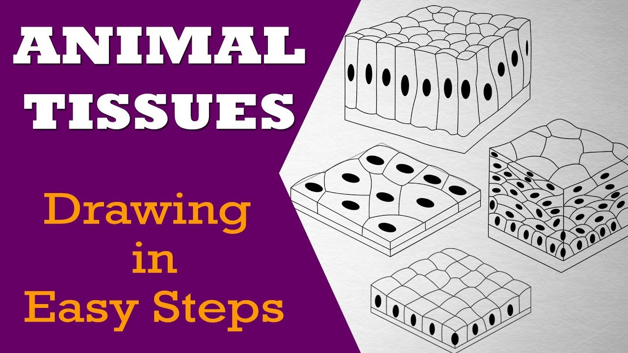
Tissue Drawing at Explore collection of Tissue Drawing

Day 18 Drawing of a tissue box with light. Medium Black Conté Crayon

HOW TO DRAW A CUTE TISSUE, STEP BY STEP

epithelial tissue, drawing Stock Image C015/2525 Science Photo

How to Draw Tissue box Step by Step YouTube
How to Draw a Tissue Box Step by Step Easy Drawing Guides Drawing
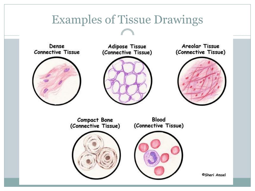
PPT Body Tissues PowerPoint Presentation, free download ID3057525
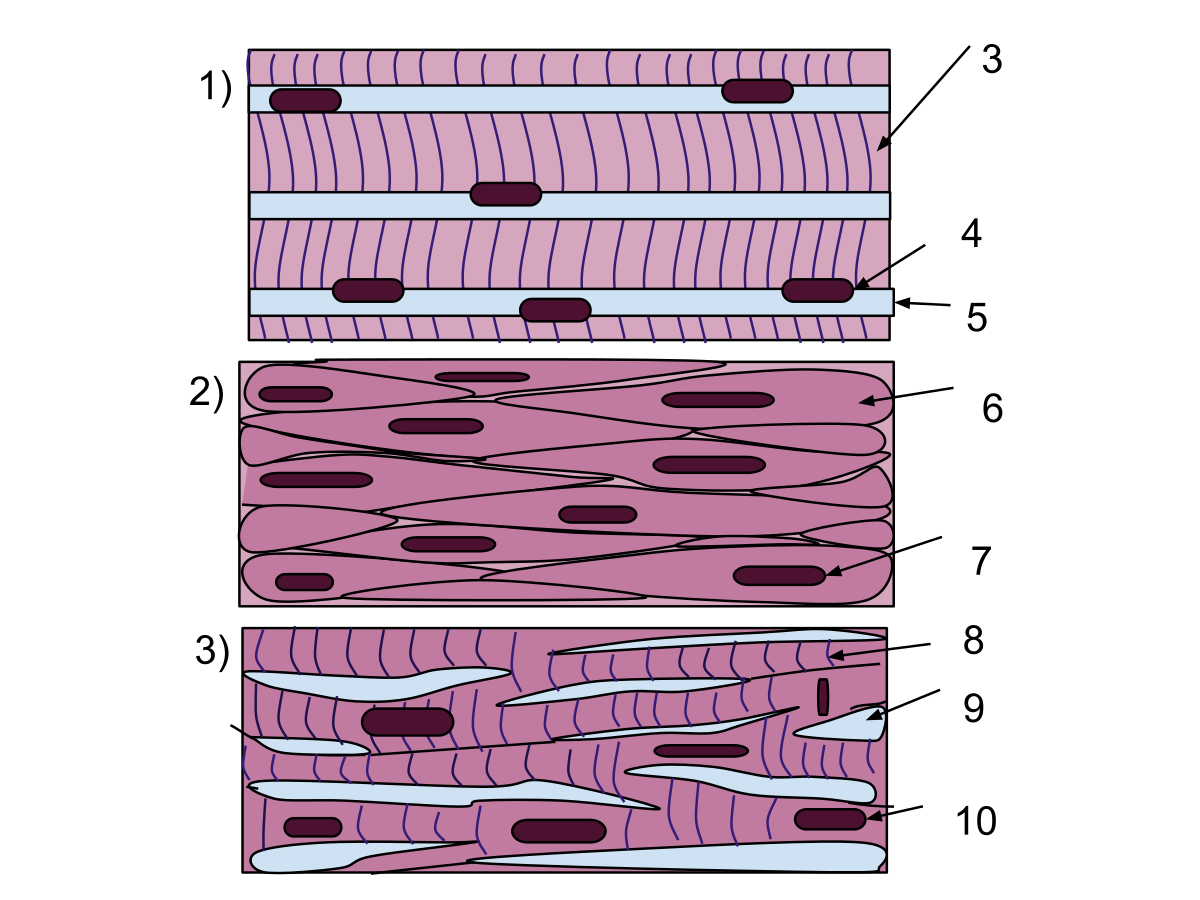
Muscle Tissue Drawing at GetDrawings Free download

How to Draw a Tissue Box 6 Steps (with Pictures) wikiHow
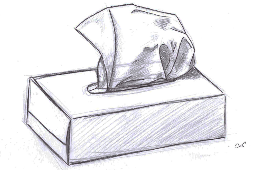
Tissue Drawing at Explore collection of Tissue Drawing
The Word Tissue Comes From A Form Of An Old French Verb Meaning “To Weave”.
Be Able To Correlate Different Types Of Epithelia With Their Locations And Functions.
Web The Term Tissue Is Used To Describe A Group Of Cells Found Together In The Body.
Microscopic Observation Reveals That The Cells In A Tissue Share Morphological Features And Are Arranged In An Orderly Pattern That Achieves The Tissue’s Functions.
Related Post: