Spine Innervation Chart
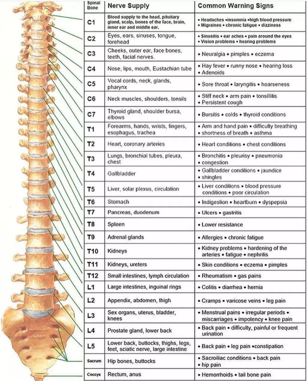
Spine Innervation Chart - Web the spine’s four sections, from top to bottom, are the cervical (neck), thoracic (abdomen,) lumbar (lower back), and sacral (toward tailbone). Thoracic spinal nerves are not part of any plexus, but give rise to the intercostal nerves directly. Web spinal nerves are mixed nerves that emerge from the spinal cord and carry both motor and sensory information between the spinal cord and various parts of the body. The vertebral column, commonly known as the spine, spinal column, or backbone, is a flexible hollow structure through which the spinal cord runs. The spinal cord begins at the base of the brain and extends into the pelvis. Supports your upper body, distributes body weight. The thyroid is closely positioned to vital structures such as the carotid artery, jugular vein, lymphatic vessels, nerves, trachea, and esophagus. Web spine deformity vol. Web there are 5 pairs of spinal nerves in the lumbar spine, labeled l1 to l5. These are muscle movement, sensation, and autonomic functions (involuntary functions). It is important to mention that after the spinal nerves exit from the spine, they join together to form four paired clusters of. Web spinal nerves are all mixed nerves with both sensory and motor fibers. Web the spinal cord can be divided into segments according to the nerve roots that branch off from it. Printed in the usa by. Web [top] what does this mean for you? This spinal column is held in place by surrounding muscles, ligaments and tendons that act as supporting guy wires. Supports your upper body, distributes body weight. When sampling thyroid nodules, they are often in proximity to the carotid artery, jugular vein, or both. All of these bones and sections are important to. Each nerve is named after the vertebra above it. It is important to mention that after the spinal nerves exit from the spine, they join together to form four paired clusters of. Your lumbar spine supports the upper two sections of your spine — the seven vertebrae in your neck (cervical spine) and 12 vertebrae in your chest (thoracic spine). Throughout the spine, intervertebral discs made of. Web this body scientific international chart beautifully depicts the normal anatomy of the spine including anterior, lateral and posterior views show the location of atlas & axis, cervical, thoracic & lumbar vertebrae, and sacrum and coccyx and curvatures of. Instead of receiving a general treatment approach, a spine therapist specializes in treating back. Web the spine’s four sections, from top to bottom, are the cervical (neck), thoracic (abdomen,) lumbar (lower back), and sacral (toward tailbone). Web spinal nerves are mixed nerves that emerge from the spinal cord and carry both motor and sensory information between the spinal cord and various parts of the body. All of these bones and sections are important to. The spinal cord begins at the base of the brain and extends into the pelvis. Web lexington, ky / accesswire / july 29, 2024 / cardiff lexington corporation (otc pink:cdix) today announced the opening of a new location of nova ortho and spine in orlando, florida. Web what is the vertebral column. Cheerag upadhyaya | media chair. Web eric potts. Web there are 5 pairs of spinal nerves in the lumbar spine, labeled l1 to l5. All of these bones and sections are important to the spine’s ability to function properly. Web eric potts | past chair. These are muscle movement, sensation, and autonomic functions (involuntary functions). Web there are 31 pairs of spinal nerves: Web the spinal cord can be divided into segments according to the nerve roots that branch off from it. These nerves play important roles in sending messages to and from the spinal cord and cauda equina, enabling the brain to communicate with parts of the lower body. To foster the use of spinal neurosurgical methods for the treatment of diseases. There are a total of: Throughout the spine, intervertebral discs made of. Web below is a chart that outlines the main functions of each of the spine nerve roots: Web your lumbar spine has several functions, including: The vertebral column, commonly known as the spine, spinal column, or backbone, is a flexible hollow structure through which the spinal cord runs. Each of the spinal nerves carries out functions that correspond to a certain region of the body. Web learn the anatomy of the spinal nerves, including their roots, components and functions faster and more efficiently with this comprehensive article. Printed in the usa by anatomical chart company. There are a total of: It is part of the axial skeleton and. The spinal cord runs through its center. Web spine deformity vol. It comprises 33 small bones called vertebrae, which remain separated by cartilaginous intervertebral discs. There are robust enlargements of the cord at the cranial and lumbosacral regions as these areas are responsible for a significant degree of skeletal muscle innervation of the upper and lower extremities, respectively. When sampling thyroid nodules, they are often in proximity to the carotid artery, jugular vein, or both. Web the spinal cord can be divided into segments according to the nerve roots that branch off from it. Web the spinal nerves have small sensory and motor branches. Web the spine’s four sections, from top to bottom, are the cervical (neck), thoracic (abdomen,) lumbar (lower back), and sacral (toward tailbone). Web your lumbar spine has several functions, including: Your spine starts at the base of your skull (head bone) and ends at your tailbone, a part of your pelvis (the large bony structure between your abdomen and legs). Web the vertebral column (spine or backbone) is a curved structure composed of bony vertebrae that are interconnected by cartilaginous intervertebral discs. Web there are 5 pairs of spinal nerves in the lumbar spine, labeled l1 to l5. Web eric potts | past chair. Instead of receiving a general treatment approach, a spine therapist specializes in treating back and neck pain. All of these bones and sections are important to the spine’s ability to function properly. Each of these nerves branches out from the spinal cord, dividing and subdividing to form a network connecting the spinal cord to every part of the body.
View of the spinal cord and the spinal nerves Download Scientific Diagram

Lumbar Spinal Nerve Chart

Spinal Nerves Anatomical Chart Spine and Cranial Nervous System

Spinal Nerve Chart Labeled

Spinal Nerve Chart Print 5x7 Etsy Nerve anatomy, Spinal nerves

Spinal Nerve Pathway Bonati Spine Institute

Nervous System Anatomy Posters Set of 6
Spinal Nerve Function MEDizzy

Spinal Nerve Chart

Spinal Nerve Function Chart
Web The Relevant Anatomy Of The Innervation Of The Musculature Of The Back By The Spinal Nerves Is Centered Around The Lumbar Spinal Nerves, Peripheral Nerves Of The Lumbar Plexus, Spinal Cord, And Lumbar Vertebral Column.
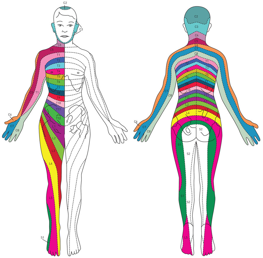
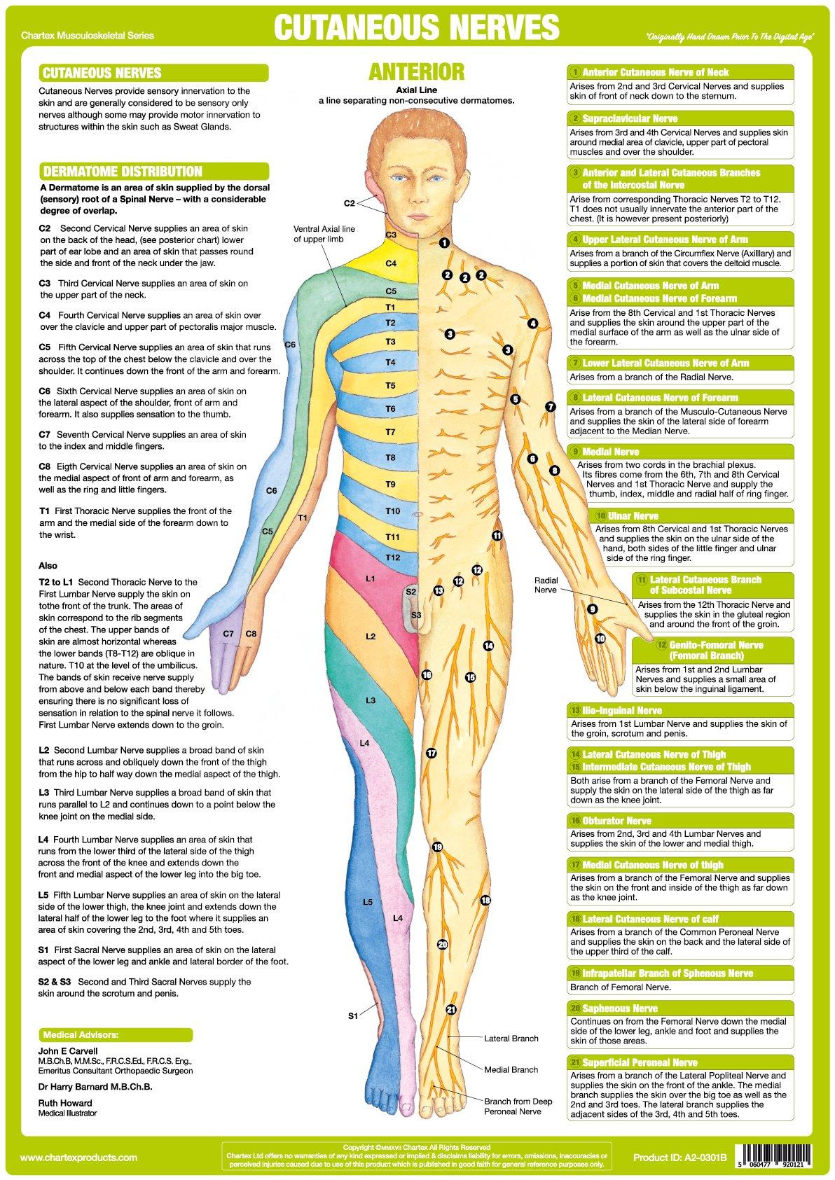
Web The Spinal Nerves Anatomical Chart Also Shows Spinal Cord Segments, Cutaneous Distribution Of Spinal Nerves And Dermal Segmentation.
Each Nerve Is Named After The Vertebra Above It.
This Spinal Column Is Held In Place By Surrounding Muscles, Ligaments And Tendons That Act As Supporting Guy Wires.
Related Post:
