Spine Chart With Nerves
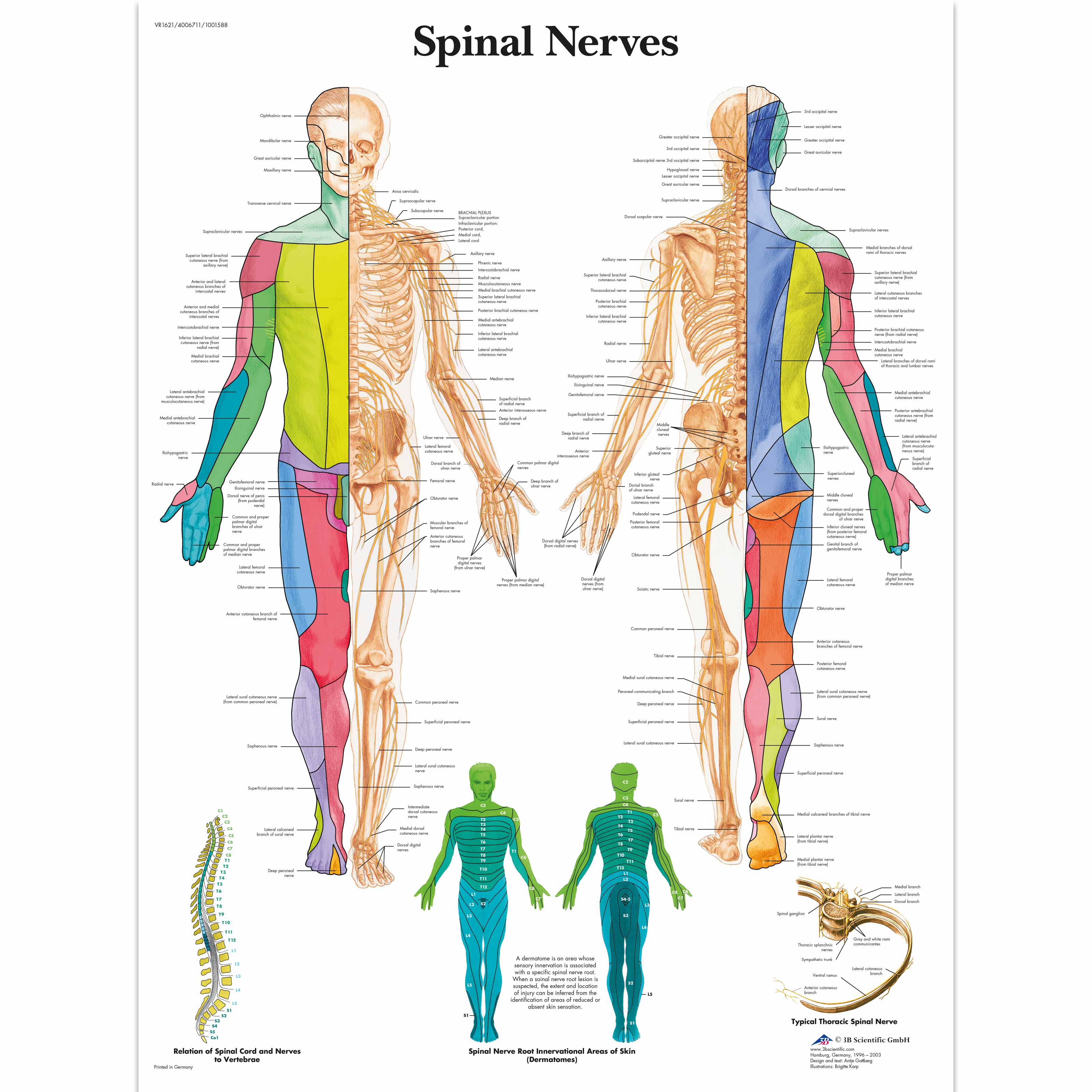
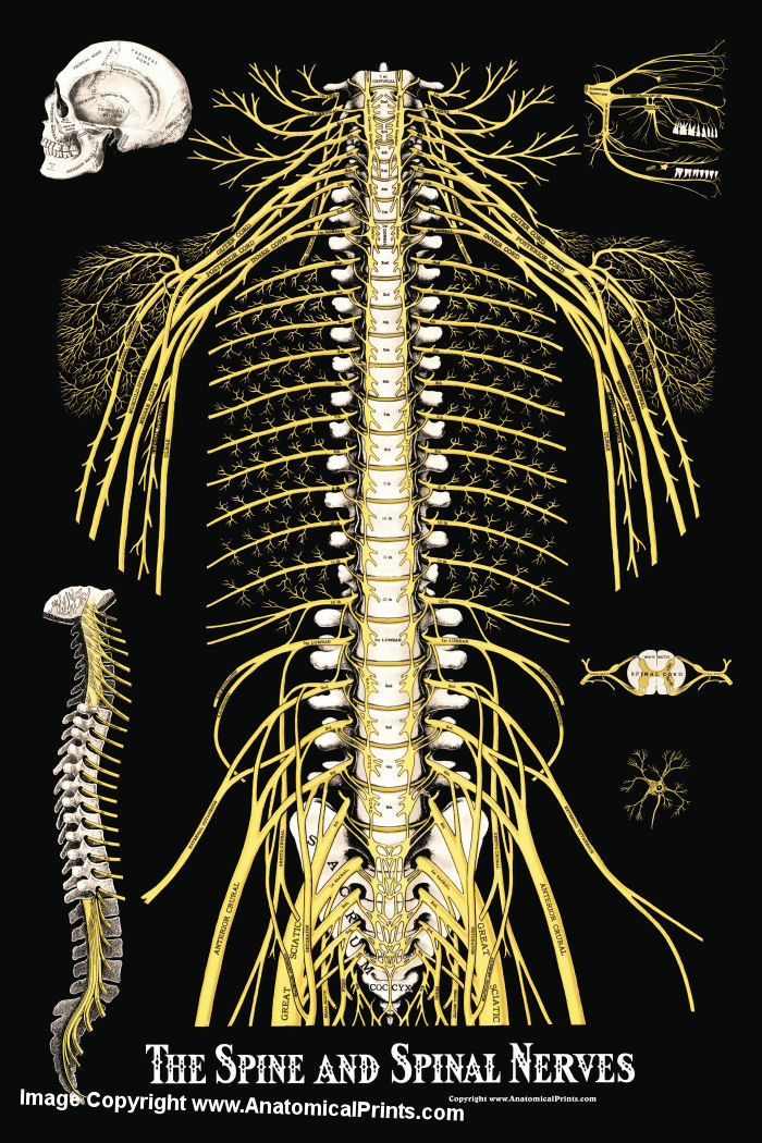
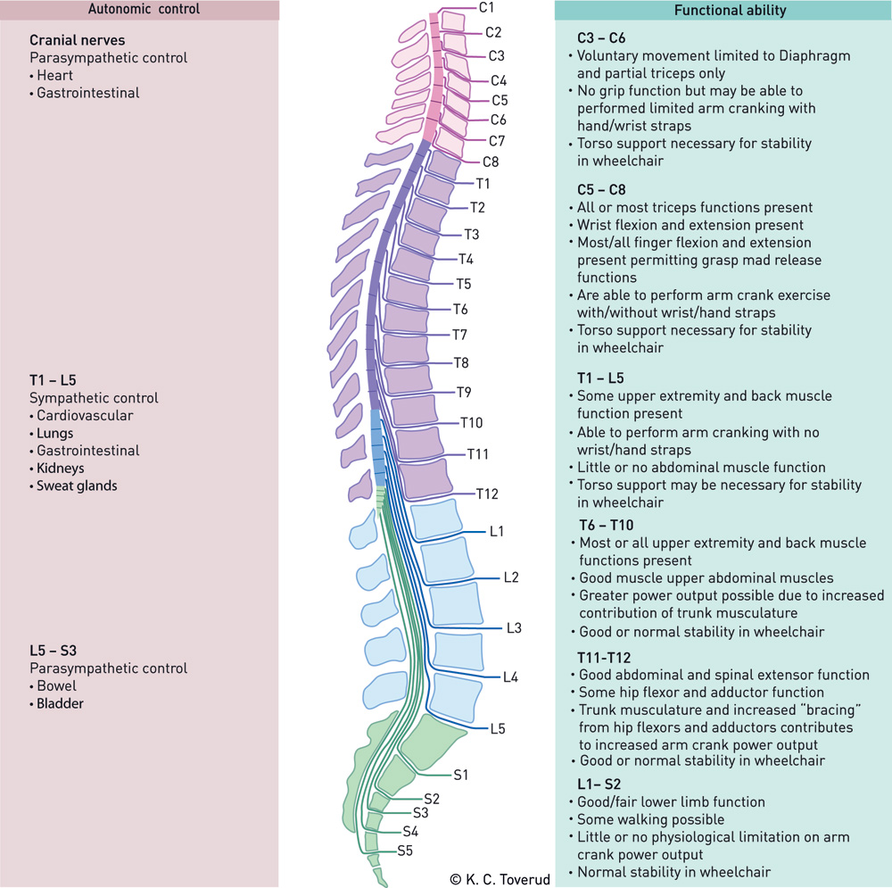
Spine Chart With Nerves - Each spinal nerve is a mixed nerve, formed from the combination of nerve root fibers from its dorsal and ventral roots. Your thoracic spine is the middle section of your spine. It is important to mention that after the spinal nerves exit from the spine, they join together to form four paired clusters of. Each spinal nerve is supplied by 2 nerve roots. These nerves are essential for transmitting sensory signals to the brain and for carrying motor commands from the brain to muscles. Your spinal cord, made up of billions of nerves, lies inside your spinal column, protected on all sides by bone. Throughout the spine, intervertebral discs made of. It consists of 12 vertebrae. 8 cervical, 12 thoracic, 5 lumbar, 5 sacral, and 1 coccygeal, named according to their corresponding vertebral levels. These nerves carry messages between your brain and muscles. Web these complex networks of nerves enable the brain to receive sensory inputs from the skin and to send motor controls for muscle movements. For the most part, the spinal nerves exit the vertebral canal through the intervertebral foramen below their corresponding vertebra. Each spinal nerve is a mixed nerve, formed from the combination of nerve root fibers from its. Web there are 31 bilateral pairs of spinal nerves, named from the vertebra they correspond to. Web spinal nerves are mixed nerves that emerge from the spinal cord and carry both motor and sensory information between the spinal cord and various parts of the body. Some tracts carry signals related to sensory information such as touch, temperature, and pain, while. Your thoracic spine is the middle section of your spine. Web spinal nerves are all mixed nerves with both sensory and motor fibers. Web there are 31 pairs of spinal nerves: Web the spinal nerves receive sensory messages from tiny nerves located in areas such as the skin, internal organs, and bones. Throughout the spine, intervertebral discs made of. Web your cervical spine — the neck area of your spine — consists of seven stacked bones called vertebrae. Some tracts carry signals related to sensory information such as touch, temperature, and pain, while others carry signals related to movement and coordination of muscles. Web these complex networks of nerves enable the brain to receive sensory inputs from the skin. Web below is a chart that outlines the main functions of each of the spine nerve roots: Your spinal cord, made up of billions of nerves, lies inside your spinal column, protected on all sides by bone. These nerves play important roles in sending messages to and from the spinal cord and cauda equina, enabling the brain to communicate with. Web how to use the spinal nerve chart: Web spinal nerves are mixed nerves that interact directly with the spinal cord to modulate motor and sensory information from the body’s periphery. Your spinal cord is a column of nerves that travels through your spinal canal. Web below is a chart that outlines the main functions of each of the spine. Other roles for the vertebrae. Spinal nerves emerge from the spinal cord and reorganize through plexuses, which then give rise to systemic nerves. Your spinal cord, made up of billions of nerves, lies inside your spinal column, protected on all sides by bone. Web nerve roots branch off from the spinal cord and exit the spinal column through small spaces. Each nerve forms from nerve fibers, known as fila radicularia, extending from the posterior (dorsal) and anterior (ventral) roots of the spinal cord. Web learn how spinal nerve roots function, and the potential symptoms of spinal nerve compression and pain in the neck and lower back. Some tracts carry signals related to sensory information such as touch, temperature, and pain,. The cord extends from your skull to your lower back. The spinal cord begins at the base of the brain and extends into the pelvis. The dorsal root is the afferent sensory root and carries sensory information to the brain. Seven in your neck (cervical spine ), 12 in your midback (thoracic spine) and 5 in your lower back (lumbar. It consists of 12 vertebrae. It is important to mention that after the spinal nerves exit from the spine, they join together to form four paired clusters of. If there is compression or irritation of these nerves due to conditions like herniated discs or bone spurs, it can result in localized or radiating pain, such as sciatica. In the cervical. Throughout the spine, intervertebral discs made of. L2, l3 and l4 spinal nerves provide sensation to the front part of your thigh and inner side of your lower leg. Web your spinal column or ‘backbone’ is made up of 24 vertebrae: Web these complex networks of nerves enable the brain to receive sensory inputs from the skin and to send motor controls for muscle movements. Each nerve forms from nerve fibers, known as fila radicularia, extending from the posterior (dorsal) and anterior (ventral) roots of the spinal cord. Web l1 spinal nerve provides sensation to your groin and genital area and helps move your hip muscles. It consists of 12 vertebrae. Each nerve is named after the vertebra above it. Each of these nerves branches out from the spinal cord, dividing and subdividing to form a network connecting the spinal cord to every part of the body. These nerves carry messages between your brain and muscles. Seven in your neck (cervical spine ), 12 in your midback (thoracic spine) and 5 in your lower back (lumbar spine). Some tracts carry signals related to sensory information such as touch, temperature, and pain, while others carry signals related to movement and coordination of muscles. The dorsal root is the afferent sensory root and carries sensory information to the brain. For the most part, the spinal nerves exit the vertebral canal through the intervertebral foramen below their corresponding vertebra. Web below is a chart that outlines the main functions of each of the spine nerve roots: It is important to mention that after the spinal nerves exit from the spine, they join together to form four paired clusters of.
Spinal Nerve Chart Print 5x7 Etsy Nerve anatomy, Spinal nerves

Printable Spinal Nerve Chart

Spinal Nerve Chart

Spinal Nerves Anatomical Chart Spine and Cranial Nervous System

Anatomical Charts and Posters Anatomy Charts Spinal Nerves Paper Chart

Printable Spinal Nerve Chart Printable World Holiday

Lumbar Spinal Nerve Chart

The Spine and Spinal Nerves Poster Clinical Charts and Supplies

Important Nerves in the Body and What They Do NorthEast Spine and

Spinal Nerve Function Anatomical Chart Anatomy Models and Anatomical
Web There Are 5 Pairs Of Spinal Nerves In The Lumbar Spine, Labeled L1 To L5.
Other Roles For The Vertebrae.
Web Spinal Nerves Are Mixed Nerves That Interact Directly With The Spinal Cord To Modulate Motor And Sensory Information From The Body’s Periphery.
Web Health Library / Body Systems & Organs / Thoracic Spine.
Related Post: