Printable Dermatome Chart
Printable Dermatome Chart - From the cells of a somite. Web clinicians can evaluate cutaneous sensation with a dermatome map as a method to localize sores within central anxious tissue, injury to particular spinal nerves, and to determine the level of the injury. These spinal sensory nerves get in the nerve root at the spine, and their branches reach to the periphery of the body. Print the dermatome map in color and at an appropriate size to ensure readability. These maps were created in the 1930s and are still frequently employed. The exact area that each dermatome covers can be different from person to person. These maps were developed in the 1930s and are frequently utilized. Web the keegan and garret map and the foerster map. Clinicians can assess cutaneous experience with a dermatome map as a way to localize sores within main anxious tissue, injury to particular back nerves, and to identify the degree of the injury. These cells differentiate into the following 3 regions: Web dermatomes help physicians to build diagrams of the spine, which aid in the diagnosis. (1) myotome, which forms some of the skeletal muscle; Web free printable dermatome chart. It is an area of skin which is innervated by the posterior (dorsal) root of a single spinal nerve. The trigeminal nerve and the maxillary nerve are among the most extensive. Web dermatomes are areas of skin, and each communicates with the brain via a single nerve. The trigeminal nerve and the maxillary nerve are among the most extensive dermatomes. Dermatome maps portray the sensory distribution of each dermatome throughout the body. These cells differentiate into the following 3 regions: Use the dermatome map as a visual aid to study the. There is actually considerable overlap between any two adjacent dermatomes. 15 13 14 st 15 tio t12 ,2,3,4 2, l5 st c6,7,8 t4 clavicles lateral parts of upper limbs medial sides of upper limbs thumb hand ring and little fingers level of nipples Web levels of principal dermatomes schematic demarcation of dermatomes shown as distinct segments. These maps were developed. Web the lists below describe locations that can be used to assess the dermatomes of the head, upper limb, torso and lower limbs. Print the dermatome map in color and at an appropriate size to ensure readability. (2) dermatome, which forms the connective tissues, including the dermis; Web dermatomes are areas of skin, and each communicates with the brain via. Web levels of principal dermatomes schematic demarcation of dermatomes shown as distinct segments. 15 13 14 st 15 tio t12 ,2,3,4 2, l5 st c6,7,8 t4 clavicles lateral parts of upper limbs medial sides of upper limbs thumb hand ring and little fingers level of nipples These cells differentiate into the following 3 regions: Clinicians can assess cutaneous experience with. These maps were developed in the 1930s and are frequently utilized. Web the dermatome map depicts the spinal nerve distribution across the body. Web the lists below describe locations that can be used to assess the dermatomes of the head, upper limb, torso and lower limbs. These spinal sensory nerves get in the nerve root at the spine, and their. There is actually considerable overlap between any two adjacent dermatomes. These spinal sensory nerves get in the nerve root at the spine, and their branches reach to the periphery of the body. It is a location of skin which is innervated by the posterior (dorsal) root of a single back nerve. These back sensory nerves get in the nerve root. Use the dermatome map as a visual aid to study the dermatomal distribution and key landmarks. Download this pdf to help you learn more about the patient's clinical condition. Web dermatomes are areas of skin, and each communicates with the brain via a single nerve. Web levels of principal dermatomes schematic demarcation of dermatomes shown as distinct segments. Dermatome maps. Web clinicians can evaluate cutaneous sensation with a dermatome map as a method to localize sores within central anxious tissue, injury to particular spinal nerves, and to determine the level of the injury. Download this pdf to help you learn more about the patient's clinical condition. Use the dermatome map as a visual aid to study the dermatomal distribution and. Web free printable dermatome chart. Clinicians can assess cutaneous experience with a dermatome map as a way to localize sores within main anxious tissue, injury to particular back nerves, and to identify the degree of the injury. Web clinicians can evaluate cutaneous sensation with a dermatome map as a method to localize sores within central anxious tissue, injury to particular. It is a location of skin which is innervated by the posterior (dorsal) root of a single back nerve. They are the keegan and garret map and the foerster map. Here, find out more about the relationship between nerves and dermatomes. Download this pdf to help you learn more about the patient's clinical condition. 1 we have also included a selection of dermatomal maps to demonstrate the region of the skin each dermatome covers. The trigeminal nerve and the maxillary nerve are among the most extensive dermatomes. Web table of contents. Web the dermatome map depicts the spinal nerve distribution across the body. “derma” implying “skin”, and “tome”, suggesting “cutting” or “thin section”. Web dermatome chart september 2018 use this dermatome chart to determine the extent of vesicular lesions that involve multiple areas. These back sensory nerves get in the nerve root at the spinal cord, and their branches reach to the periphery of the body. Dermatome maps depict the sensory distribution of each dermatome across the body. These maps were created in the 1930s and are still frequently employed. 15 13 14 st 15 tio t12 ,2,3,4 2, l5 st c6,7,8 t4 clavicles lateral parts of upper limbs medial sides of upper limbs thumb hand ring and little fingers level of nipples Web dermatomes are areas of skin, and each communicates with the brain via a single nerve. Web clinicians can evaluate cutaneous sensation with a dermatome map as a method to localize sores within main anxious tissue, injury to particular spinal nerves, and to identify the extent of the injury.
Reference Chart Dermatomes
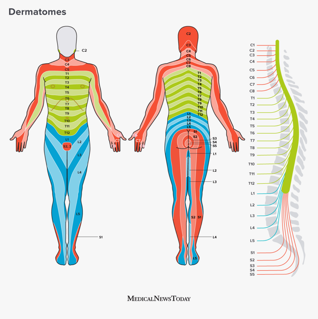
Dermatomes Chart Arm Printable Lab

Printable Dermatome Chart
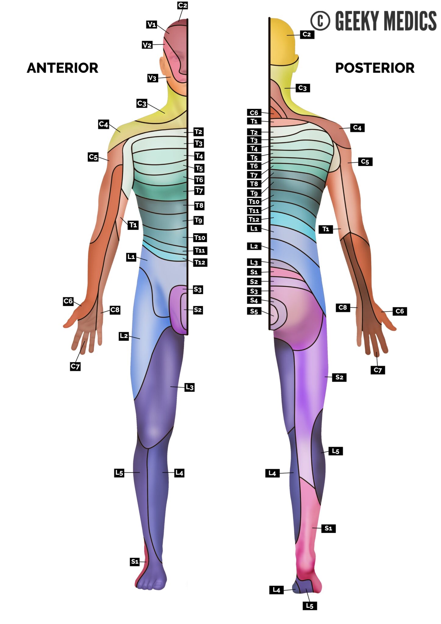
Dermatomes Upper Limb

Printable Dermatome Chart
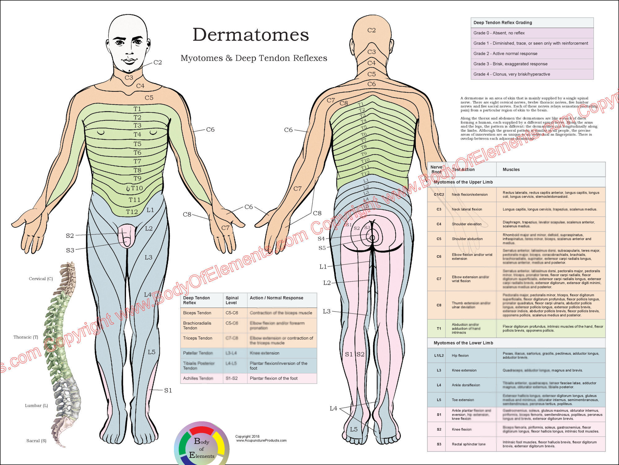
Printable Dermatome Chart
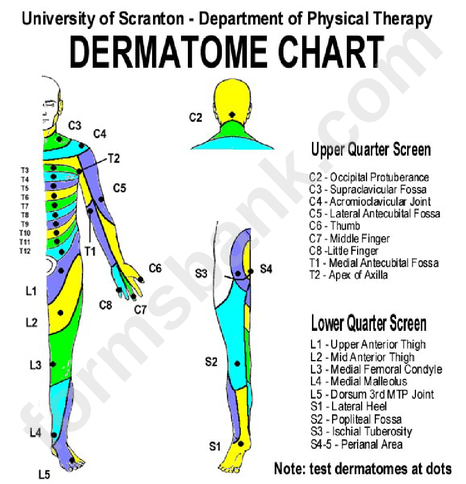
Dermatome Chart printable pdf download
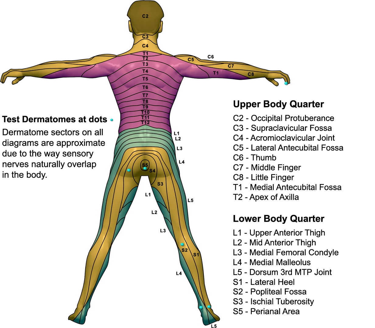
Dermatomes And Myotomes Anatomy Geeky Medics Adams Printable Map
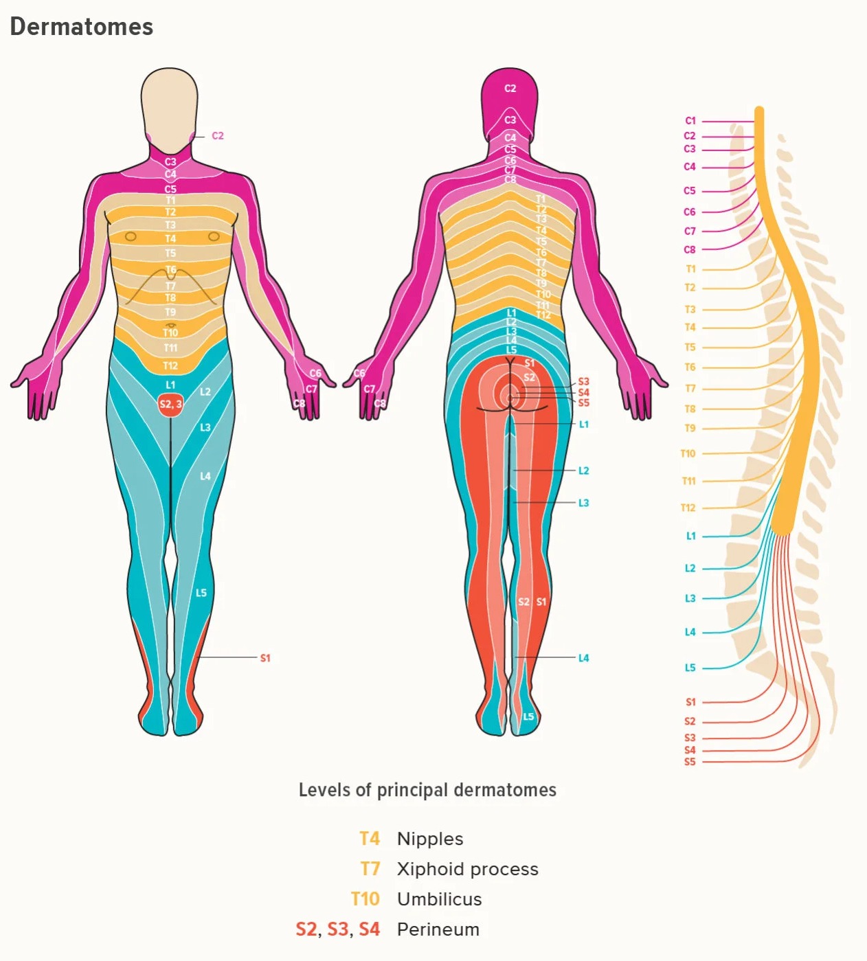
Printable Dermatome Map Printable JD

Dermatomes Back Chart
Web The Keegan And Garret Map And The Foerster Map.
“Derma” Implying “Skin”, And “Tome”, Implying “Cutting” Or “Thin Section”.
Trigeminal Nerve (Cn V) V1:
(1) Myotome, Which Forms Some Of The Skeletal Muscle;
Related Post: