Pineal Cyst Size Chart
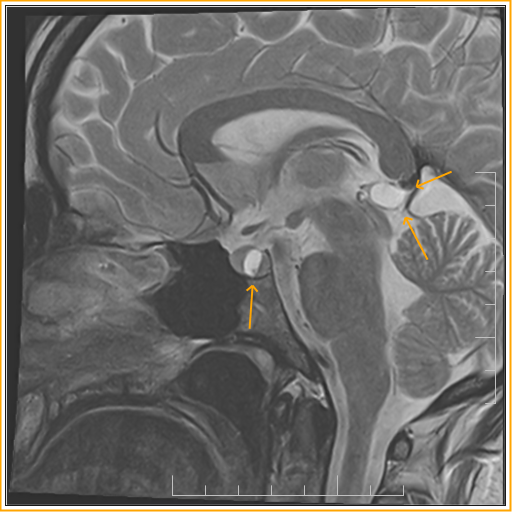
Pineal Cyst Size Chart - Web pineal cysts can be categorised on mr imaging as either simple or atypical. Pineal gland cysts are common. As many as 2 percent of healthy adults develop this kind of cyst. When larger they can present with mass effect on the tectal plate leading to compression of the superior colliculi and parinaud syndrome. Web scatter chart showing patients with pineal cyst progression and regression by age and cyst diameter While many pineal cysts are harmless and cause no symptoms, some can be problematic. If the cerebral aqueduct is compressed, they may also result in obstructive hydrocephalus. In most cases, no treatment is necessary for a pineal gland cyst. 3 simple pineal cysts are unilocular, often with a smooth, thin wall (which may or may not enhance) and can range in size from <<strong>5 mm to</strong> >25 mm in maximum diameter ( figure 1 ). When larger, they may compress the tectal plate and aqueduct resulting in hydrocephalus. Web the vast majority of pineal cysts are small (<<strong>1 cm</strong>) and asymptomatic. Web scatter chart showing patients with pineal cyst progression and regression by age and cyst diameter When larger, they may compress the tectal plate and aqueduct resulting in hydrocephalus. Sometimes an mri of the pineal cyst needs to be repeated with an intravenous contrast (dye) to rule. 3 simple pineal cysts are unilocular, often with a smooth, thin wall (which may or may not enhance) and can range in size from <<strong>5 mm to</strong> >25 mm in maximum diameter ( figure 1 ). These cysts are benign, which means not malignant or cancerous. Web pineal cysts can be categorised on mr imaging as either simple or atypical.. If the cerebral aqueduct is compressed, they may also result in obstructive hydrocephalus. When larger they can present with mass effect on the tectal plate leading to compression of the superior colliculi and parinaud syndrome. Rarely does a pineal gland cyst cause headaches or any other symptoms. While many pineal cysts are harmless and cause no symptoms, some can be. Web the vast majority of pineal cysts are small (<<strong>1 cm</strong>) and asymptomatic. When larger they can present with mass effect on the tectal plate leading to compression of the superior colliculi and parinaud syndrome. While many pineal cysts are harmless and cause no symptoms, some can be problematic. Rarely does a pineal gland cyst cause headaches or any other. When larger, they may compress the tectal plate and aqueduct resulting in hydrocephalus. Rarely does a pineal gland cyst cause headaches or any other symptoms. When larger they can present with mass effect on the tectal plate leading to compression of the superior colliculi and parinaud syndrome. As many as 2 percent of healthy adults develop this kind of cyst.. Web the vast majority of pineal cysts are small (<<strong>1 cm</strong>) and asymptomatic. Web scatter chart showing patients with pineal cyst progression and regression by age and cyst diameter While many pineal cysts are harmless and cause no symptoms, some can be problematic. 3 simple pineal cysts are unilocular, often with a smooth, thin wall (which may or may not. When larger, they may compress the tectal plate and aqueduct resulting in hydrocephalus. In most cases, no treatment is necessary for a pineal gland cyst. As many as 2 percent of healthy adults develop this kind of cyst. While many pineal cysts are harmless and cause no symptoms, some can be problematic. Pineal gland cysts are common. If the cerebral aqueduct is compressed, they may also result in obstructive hydrocephalus. While many pineal cysts are harmless and cause no symptoms, some can be problematic. Rarely does a pineal gland cyst cause headaches or any other symptoms. Pineal gland cysts are common. In most cases, no treatment is necessary for a pineal gland cyst. These cysts are benign, which means not malignant or cancerous. If the cerebral aqueduct is compressed, they may also result in obstructive hydrocephalus. Rarely does a pineal gland cyst cause headaches or any other symptoms. When larger, they may compress the tectal plate and aqueduct resulting in hydrocephalus. Web scatter chart showing patients with pineal cyst progression and regression by. Web the vast majority of pineal cysts are small (<<strong>1 cm</strong>) and asymptomatic. Sometimes an mri of the pineal cyst needs to be repeated with an intravenous contrast (dye) to rule out a pineal tumor. Rarely does a pineal gland cyst cause headaches or any other symptoms. As many as 2 percent of healthy adults develop this kind of cyst.. In most cases, no treatment is necessary for a pineal gland cyst. These cysts are benign, which means not malignant or cancerous. As many as 2 percent of healthy adults develop this kind of cyst. Sometimes an mri of the pineal cyst needs to be repeated with an intravenous contrast (dye) to rule out a pineal tumor. Web the vast majority of pineal cysts are small (<1 cm) and asymptomatic. If the cerebral aqueduct is compressed, they may also result in obstructive hydrocephalus. When larger they can present with mass effect on the tectal plate leading to compression of the superior colliculi and parinaud syndrome. When larger, they may compress the tectal plate and aqueduct resulting in hydrocephalus. Web pineal cysts are common and generally less than 15 mm in greatest dimension. Rarely does a pineal gland cyst cause headaches or any other symptoms. Pineal gland cysts are common. 3 simple pineal cysts are unilocular, often with a smooth, thin wall (which may or may not enhance) and can range in size from <<strong>5 mm to</strong> >25 mm in maximum diameter ( figure 1 ).
Pineal Cyst Size Chart

Pineal Cyst Simulating Pinealoblastoma in 11 Children With

Pineal Cyst Size Chart

Pineal cysts diameters across the surgical criteria groups (a

Pineal Cyst Size Chart

Brain Cyst Size Chart

Pineal Cyst Simulating Pinealoblastoma in 11 Children With

Pineal cysts diameters across the surgical criteria groups (a

Pineal cysts diameters across the surgical criteria groups (a

Pineal Cyst Size Chart
While Many Pineal Cysts Are Harmless And Cause No Symptoms, Some Can Be Problematic.
Web Scatter Chart Showing Patients With Pineal Cyst Progression And Regression By Age And Cyst Diameter
Web Pineal Cysts Can Be Categorised On Mr Imaging As Either Simple Or Atypical.
Related Post: