Medical Drawing Of Heart
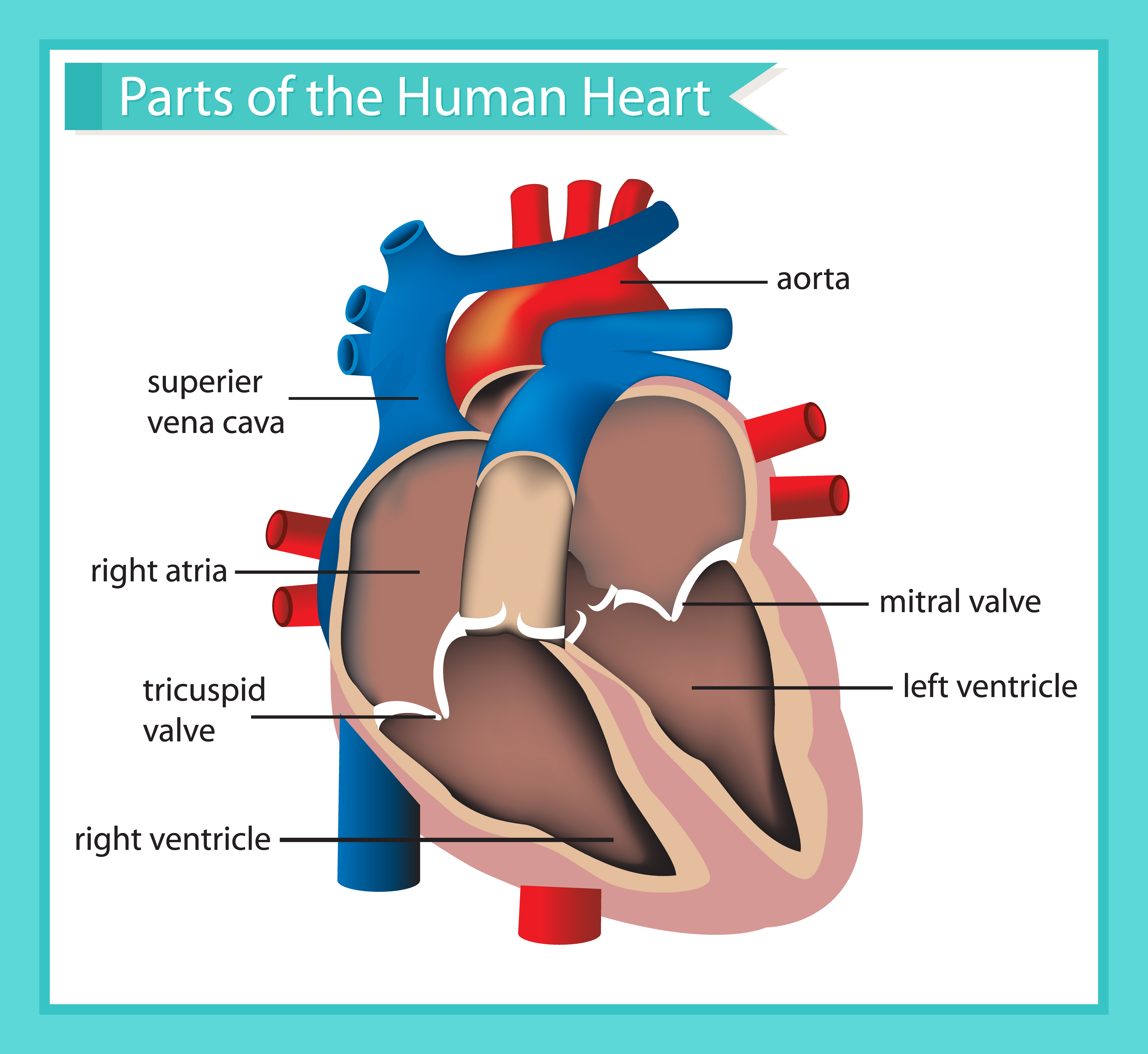
Medical Drawing Of Heart - Titles include heart disease, hypertension, target heart rate and more. Find a piece of paper and something to draw with. There is also a murmurs tab that includes some of the more common murmurs we come across. Web heart, organ that serves as a pump to circulate the blood. Web these anatomical heart medical illustrations are highly detailed drawings that blend art with science. Blood flows through the heart. Web this video shows you how to draw a heart for your scientific figure and graphical abstract. Xxxl very detailed human heart. Web choose from normal, or abnormal anatomy illustrations in a variety of sizes. Our extensive library features carefully curated medical visuals, perfect for creating compelling educational aids and enriching your research work. View anatomical heart drawing videos. 48k views 1 year ago cardiovascular system. 🎨 drawbiomed is a channel for scientists to learn professional scientific illustrations for their. Browse 2,244 anatomical heart drawing photos and images available, or start a new search to explore more photos and images. This week, the royal foundation centre for early. The inferior tip of the heart, known as the apex, rests just superior to the diaphragm. Web postprandial blood sugar is the level of glucose in your blood after eating. You can use these drawings for patient education, teaching students and as a reference for seasoned practitioners in their medical offices. Web the intricate anatomy of the heart can be. Find a piece of paper and something to draw with. It tends to spike one hour after eating and normalize one hour later. You can use these drawings for patient education, teaching students and as a reference for seasoned practitioners in their medical offices. This week, the royal foundation centre for early. Balancing daily responsibilities with happiness is indeed an. It also takes away carbon dioxide and other waste so other organs can dispose of them. Blood flows through the heart. You can use these drawings for patient education, teaching students and as a reference for seasoned practitioners in their medical offices. Web how it works. Web anatomy of the human heart and coronaries: It consists of four main chambers: Find a piece of paper and something to draw with. Web the heart is a muscular organ that pumps blood around the body by circulating it through the circulatory/vascular system. Web your heart’s main function is to move blood throughout your body. View anatomical heart drawing videos. The inferior tip of the heart, known as the apex, rests just superior to the diaphragm. Xxxl very detailed human heart. This tool provides access to several medical illustrations, allowing the user to interactively discover heart anatomy. 48k views 1 year ago cardiovascular system. Right lung, left lung, heart copyright american heart association download (218.4 kb) Web this video shows you how to draw a heart for your scientific figure and graphical abstract. The superior vena cava and inferior vena cava (not shown) bring deoxygenated blood from the body into the right atrium, from which it enters the right ventricle. Web the heart is a mostly hollow, muscular organ composed of cardiac muscles and connective tissue. They will be to the lower left of the aorta. In this lecture, dr mike shows the two best ways to draw and. Blood brings oxygen and nutrients to your cells. 48k views 1 year ago cardiovascular system. It consists of four main chambers: The pulmonary trunk delivers deoxygenated blood to the lungs. The heart is an amazing organ. Web the heart is a muscular organ that pumps blood around the body by circulating it through the circulatory/vascular system. How to visualize anatomic structures. It also takes away carbon dioxide and other waste so other organs can dispose of them. 48k views 1 year ago cardiovascular system. Web the heart is a mostly hollow, muscular organ composed of cardiac muscles and connective tissue that acts as a pump to distribute blood throughout the body’s tissues. Find a piece of paper and something to draw with. There are two of them. Titles include heart disease, hypertension, target heart rate and more. Web these anatomical heart medical illustrations are highly detailed drawings that blend art with science. The superior vena cava and inferior vena cava (not shown) bring deoxygenated blood from the body into the right atrium, from which it enters the right ventricle. To find a good diagram, go to google images, and type in the internal structure of the human heart. Find a piece of paper and something to draw with. It splits into the right and left pulmonary. Images are labelled, providing an invaluable medical and anatomical tool. The open heart healing podcast is an excellent resource for anyone seeking a bit of inspiration and guidance in navigating life's ups and downs. Web princess kate ’s newest initiative is supporting organizations that are making resources easily accessible to families across the united kingdom. On its superior end, the base of the heart is attached to the aorta, pulmonary arteries and veins, and the vena cava. Web this cross section of the heart shows the right ventricle, tricuspid valve, left ventricle, bicuspid (mitral) valve, left atrium, right atrium, superior vena cava, inferior vena cava, aorta, aortic valve, papillary muscle, chordae tendineae, and trabeculae carneae. Web postprandial blood sugar is the level of glucose in your blood after eating. The pulmonary trunk delivers deoxygenated blood to the lungs. Postprandial blood sugar can be measured with a postprandial glucose (ppg) test to determine if you have prediabetes (140 to 199 mg/dl), type 2 diabetes (200 mg/dl and over), or gestational. Web dr matt & dr mike. Find an image that displays the entire heart, and click on it to enlarge it. Start with the pulmonary veins.
Antique medical scientific illustration highresolution heart Human

Anatomical illustration by Elisa Schorn Medical illustration

Scientific medical illustration of parts of the human heart 685453

8 Anatomical Heart Drawings! Anatomical heart drawing, Heart drawing

Heart Health Free Stock Photo Medical illustration of a human heart

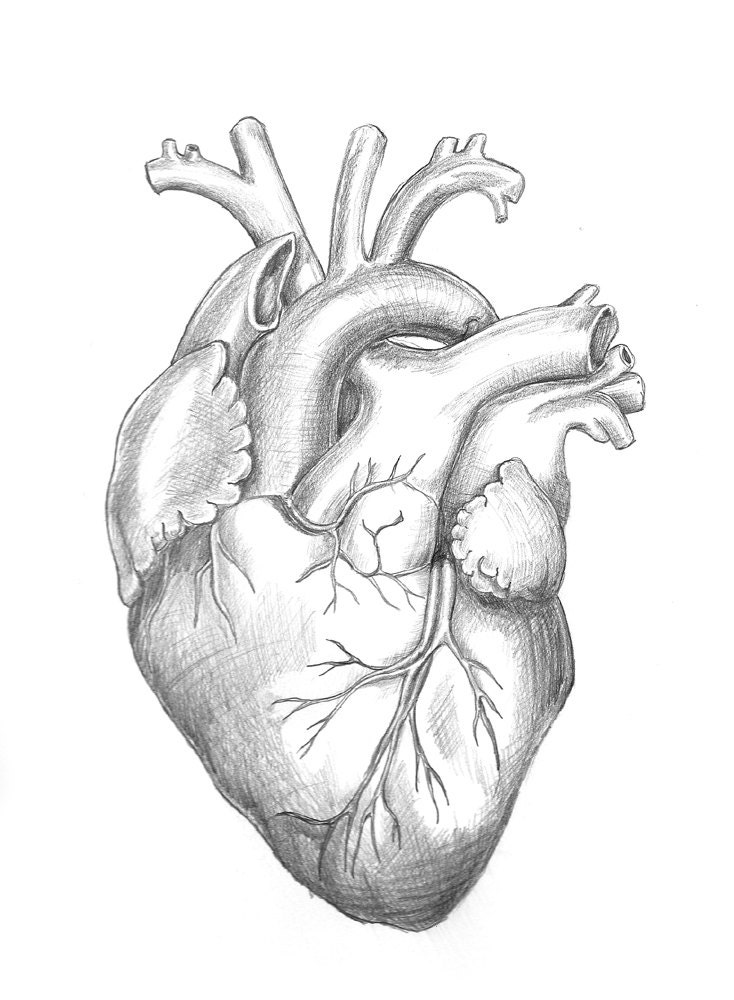
Anatomy Heart Original Unframed Pencil Drawing

Human Heart by Tom Connell at

Heart Human Anatomy sketch vector illustration 10810706 Vector Art at

Anatomical Medical Illustration, Human Heart Organ Illustration Stock

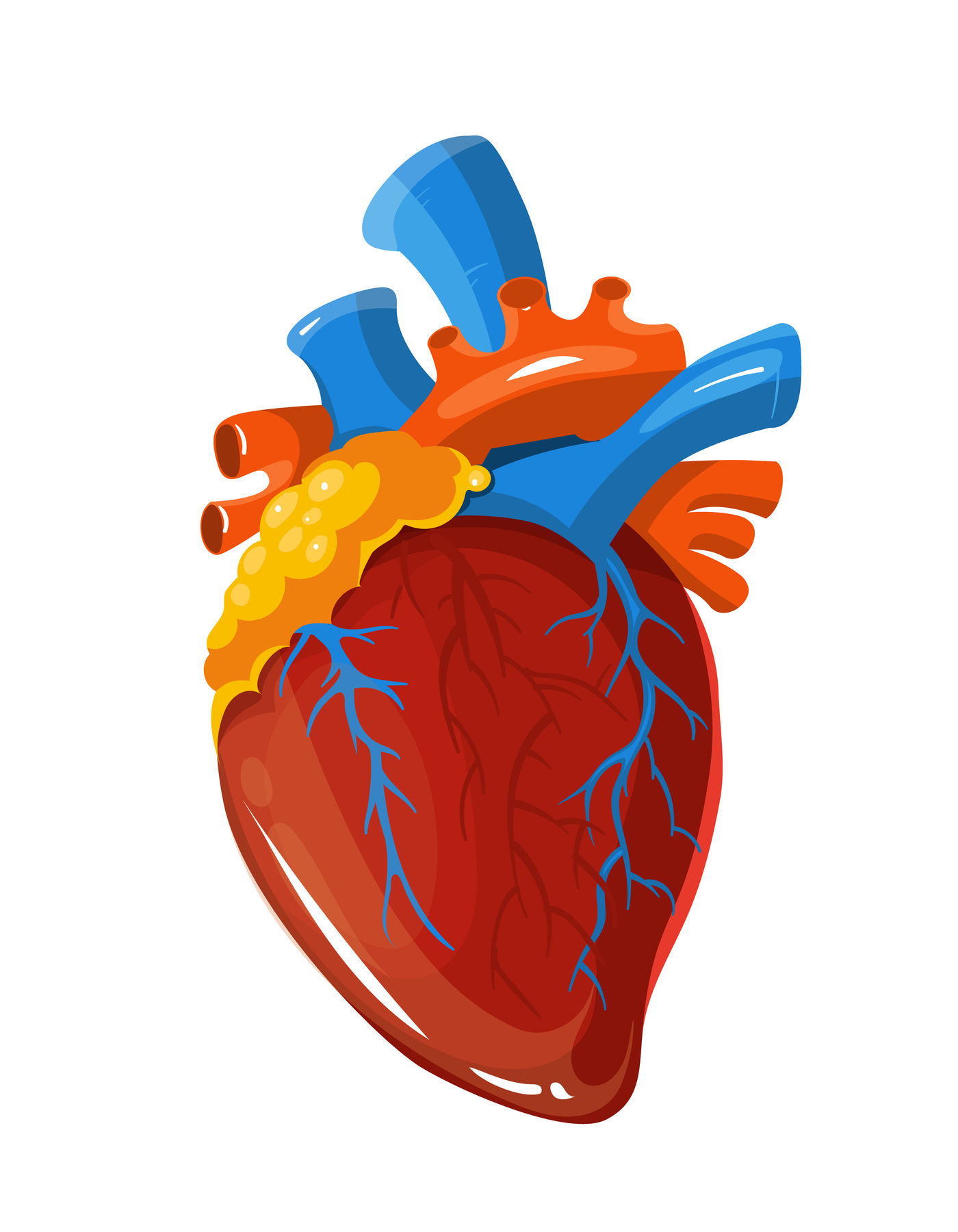
Human heart anatomy vector medical illustration By Microvector
Web Your Heart Sure Does Work Hard, But That Doesn’t Mean You Have To Work Hard To Draw It!
It Tends To Spike One Hour After Eating And Normalize One Hour Later.
Crochet Art And Funny Cats Are Wholesome, But There's A Weird Side To The Internet, Too.
Blood Leaves The Right Ventricle Via The Pulmonary Trunk.
Related Post: