Lv Strain Pattern
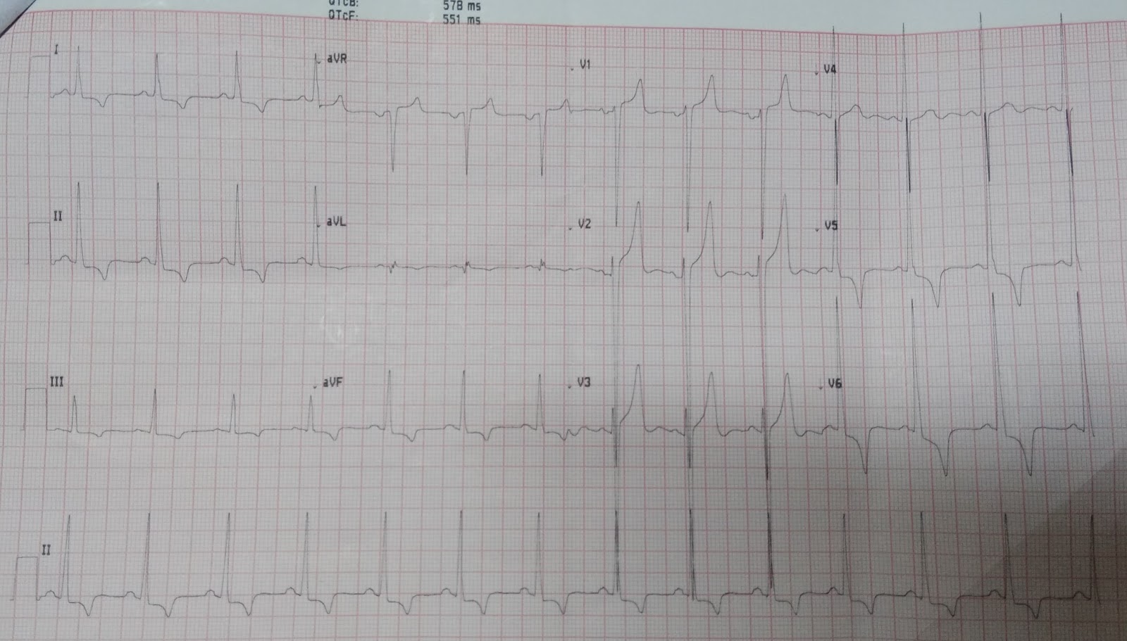
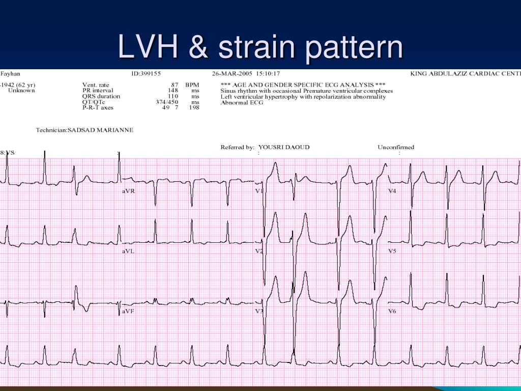
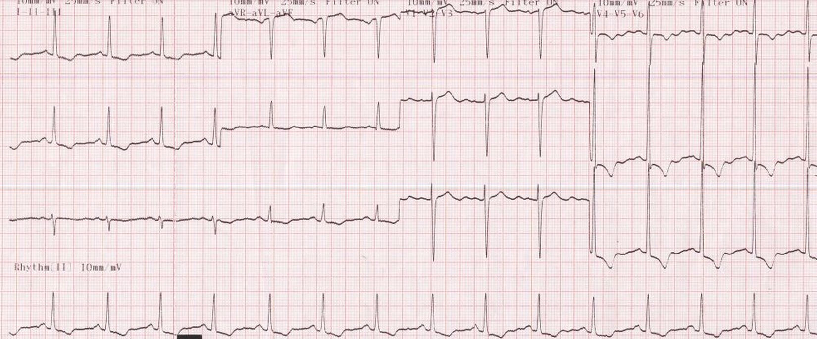
Lv Strain Pattern - Web myocardial strain is a dimensionless variable representing the change in length between two points over the cardiac cycle, and can be quantified using echocardiography or cmr tissue tracking. There will also be slight st elevations (reciprocal to the depressions) in leads with negative qrss. Web with the advent of left ventricular deformation (strain) analysis, a new and robust means for assessing left ventricular function has emerged. Sensation of rapid, fluttering or pounding heartbeats, called palpitations; Shortness of breath, especially while lying down; Contemporary research and guidelines have attempted to standardize the definition, acquisition and measurement of left ventricular strain. Web ecg changes in left ventricular hypertrophy (lvh) and right ventricular hypertrophy (rvh). Web left ventricular hypertrophy (lvh) refers to an increase in the size of myocardial fibers in the main cardiac pumping chamber. These differences were statistically significant. The two most common pressure overload states are systemic hypertension and aortic stenosis. The two most common pressure overload states are systemic hypertension and aortic stenosis. This is seen in sloping st depressions in all leads with upright qrs complexes. Web myocardial strain is a dimensionless variable representing the change in length between two points over the cardiac cycle, and can be quantified using echocardiography or cmr tissue tracking. These differences were statistically. Chest pain, often when exercising; But symptoms may occur as the strain on the heart worsens. Sensation of rapid, fluttering or pounding heartbeats, called palpitations; Contemporary research and guidelines have attempted to standardize the definition, acquisition and measurement of left ventricular strain. Web when lvh is caused by a pathological condition, we often see the strain pattern, which is st. Web left ventricular hypertrophy (lvh): Web myocardial strain is a dimensionless variable representing the change in length between two points over the cardiac cycle, and can be quantified using echocardiography or cmr tissue tracking. Shortness of breath, especially while lying down; The two most common pressure overload states are systemic hypertension and aortic stenosis. These differences were statistically significant. Huge precordial r and s waves that overlap with the adjacent leads (sv2 + rv6 >> 35 mm). Shortness of breath, especially while lying down; The two most common pressure overload states are systemic hypertension and aortic stenosis. Web in order to diagnose lvh from the ecg, we must also show repolarization abnormalities, called the strain pattern. There will also. Such hypertrophy is usually the response to a chronic pressure or volume load. Web ecg changes in left ventricular hypertrophy (lvh) and right ventricular hypertrophy (rvh). But symptoms may occur as the strain on the heart worsens. Web when lvh is caused by a pathological condition, we often see the strain pattern, which is st depression and t wave inversion. This is seen in sloping st depressions in all leads with upright qrs complexes. Web left ventricular hypertrophy itself doesn't cause symptoms. Shortness of breath, especially while lying down; Web when lvh is caused by a pathological condition, we often see the strain pattern, which is st depression and t wave inversion in leads with upright qrs complexes (the lateral. Web myocardial strain is a dimensionless variable representing the change in length between two points over the cardiac cycle, and can be quantified using echocardiography or cmr tissue tracking. Web ecg changes in left ventricular hypertrophy (lvh) and right ventricular hypertrophy (rvh). Web left ventricular hypertrophy (lvh) refers to an increase in the size of myocardial fibers in the main. Web when lvh is caused by a pathological condition, we often see the strain pattern, which is st depression and t wave inversion in leads with upright qrs complexes (the lateral leads). Web with the advent of left ventricular deformation (strain) analysis, a new and robust means for assessing left ventricular function has emerged. These differences were statistically significant. But. Web left ventricular hypertrophy (lvh): Web myocardial strain is a dimensionless variable representing the change in length between two points over the cardiac cycle, and can be quantified using echocardiography or cmr tissue tracking. Sensation of rapid, fluttering or pounding heartbeats, called palpitations; But symptoms may occur as the strain on the heart worsens. Such hypertrophy is usually the response. Such hypertrophy is usually the response to a chronic pressure or volume load. Web when lvh is caused by a pathological condition, we often see the strain pattern, which is st depression and t wave inversion in leads with upright qrs complexes (the lateral leads). These differences were statistically significant. But symptoms may occur as the strain on the heart. Huge precordial r and s waves that overlap with the adjacent leads (sv2 + rv6 >> 35 mm). But symptoms may occur as the strain on the heart worsens. Web left ventricular hypertrophy itself doesn't cause symptoms. Shortness of breath, especially while lying down; Web when lvh is caused by a pathological condition, we often see the strain pattern, which is st depression and t wave inversion in leads with upright qrs complexes (the lateral leads). Web left ventricular hypertrophy (lvh) refers to an increase in the size of myocardial fibers in the main cardiac pumping chamber. Web with the advent of left ventricular deformation (strain) analysis, a new and robust means for assessing left ventricular function has emerged. Web left ventricular hypertrophy (lvh): There will also be slight st elevations (reciprocal to the depressions) in leads with negative qrss. Web ecg changes in left ventricular hypertrophy (lvh) and right ventricular hypertrophy (rvh). Chest pain, often when exercising; Sensation of rapid, fluttering or pounding heartbeats, called palpitations; These differences were statistically significant. Fainting or a feeling of. This is seen in sloping st depressions in all leads with upright qrs complexes. Such hypertrophy is usually the response to a chronic pressure or volume load..jpg)
ECG Interpretation ECG Interpretation Review 51 (Chamber Enlargement

Lv Strain Values IUCN Water

Dr. Smith's ECG Blog Syncope and ST Elevation on the Prehospital ECG

How to differentiate LV strain pattern from primary LV ischemia ? Dr

Cardiology window Left ventricular hypertrophy with LV strain pattern

PPT ECG PRACTICAL APPROACH PowerPoint Presentation, free download

The ECG in left ventricular hypertrophy (LVH) criteria and

Left Ventricular Hypertrophy LVH with Strain Pattern on ECG YouTube

Patterns of Left Ventricular Longitudinal Strain and Strain Rate in

Left ventricular hypertrophy (LVH) with strain pattern
The Two Most Common Pressure Overload States Are Systemic Hypertension And Aortic Stenosis.
Web Myocardial Strain Is A Dimensionless Variable Representing The Change In Length Between Two Points Over The Cardiac Cycle, And Can Be Quantified Using Echocardiography Or Cmr Tissue Tracking.
Web In Order To Diagnose Lvh From The Ecg, We Must Also Show Repolarization Abnormalities, Called The Strain Pattern.
Contemporary Research And Guidelines Have Attempted To Standardize The Definition, Acquisition And Measurement Of Left Ventricular Strain.
Related Post: