Knee Anatomy Drawing
Knee Anatomy Drawing - Learn about the muscles, tendons, bones, and ligaments that comprise the knee joint anatomy. Web in this episode of simplified constructive anatomy, we cover the structure of the legs and knees. Web the anatomy of the knee consists of 3 main bones: The knee is the joint in the middle of your leg. What does the knee joint do? Finally, draw in the hamstrings covering the calves at the back of the knee. The main function of this joint is flexion and extension of the lower leg. The femur, tibia and patella. When drawing a knee, we have two big volumes—the upper and lower leg. Web the knee is a complex joint that flexes, extends, and twists slightly from side to side. It is formed by articulations between the patella, femur and tibia. These three bones are covered in articular cartilage which is an extremely hard, smooth substance designed to decrease the friction forces. Web the knee is a complex joint that flexes, extends, and twists slightly from side to side. Knee and elbow vector illustration. Web how to draw knees. While it's already relatively simple, we can still study the bones and joints, and simplify. Web knee joint anatomy consists of muscles, ligaments, cartilage and tendons. Front view of normal knee joint. Web the knee joint is the junction of the thigh and leg. Web this article takes a concise look at the anatomy of the knee joint and describes. Web in this video lesson, you will discover how to draw knees from life with the necessary knowledge of this joint's proportions and anatomy. When drawing a knee, we have two big volumes—the upper and lower leg. This connection of the femur and tibia is a joint called the tibiofemoral joint. Web before you start drawing knees, it is essential. Web how to draw knees. Knee joint icon gray illustration on white. When drawing a knee, we have two big volumes—the upper and lower leg. This connection of the femur and tibia is a joint called the tibiofemoral joint. The muscles that affect the knee’s movement run along the thigh and calf. Stabilizing you and helping keep your balance. Adding in the appropriate level of detail depending on the style of your illustration. While it's already relatively simple, we can still study the bones and joints, and simplify. The knee joint is made up of three bones: Then draw the quadriceps muscles, and indicate the patella and its tendon down to the. Your knees have several important jobs, including: Where is the knee joint located? The femur, tibia, and patella. Web the anatomy of the knee consists of 3 main bones: Knee joint icon gray illustration on white. The muscles that affect the knee’s movement run along the thigh and calf. Web let us examine the knee anatomy. When drawing a knee, we have two big volumes—the upper and lower leg. Two rounded, convex processes (known as condyles) on the distal end of the femur meet two rounded, concave condyles at the proximal end of the tibia. The. Web the knee is a complex joint that flexes, extends, and twists slightly from side to side. Web this article takes a concise look at the anatomy of the knee joint and describes the processes and conditions that cause pain in the different aspects (parts) of the knee. Two rounded, convex processes (known as condyles) on the distal end of. Web establish proportions and angles with skeletal guidelines, then work on identifying and drawing the rhythms of the shape, then fill in the muscle groups, and finally rework the overall drawing correcting for any errors. The thigh bone ( femur ), the shin bone ( tibia) and the kneecap ( patella) articulate through tibiofemoral and patellofemoral joints. Knee joint icon. Web how to draw knees. Web in this video lesson, you will discover how to draw knees from life with the necessary knowledge of this joint's proportions and anatomy. Knee joint icon gray illustration on white. Where is the knee joint located? Learn about the muscles, tendons, bones, and ligaments that comprise the knee joint anatomy. What does the knee joint do? Using basic shapes to create the general silhouette of the figure. The femur and the tibia are the main movers of the joint to allow for the hinge motion. Building the muscle structure and anatomy on top of those shapes. Web in this video lesson, you will discover how to draw knees from life with the necessary knowledge of this joint's proportions and anatomy. Your knees have several important jobs, including: Web before you start drawing knees, it is essential to understand the anatomy of the knee joint. Web the knee joint is the junction of the thigh and leg. Each anatomical structure was labeled interactively. Web the knee is a complex joint that flexes, extends, and twists slightly from side to side. The structure of a normal knee joint. Knee and elbow vector illustration. They are attached to the femur (thighbone), tibia (shinbone), and fibula (calf bone) by fibrous tissues called. While it's already relatively simple, we can still study the bones and joints, and simplify. The thigh bone ( femur ), the shin bone ( tibia) and the kneecap ( patella) articulate through tibiofemoral and patellofemoral joints. Web the knee joint is a synovial joint that connects three bones;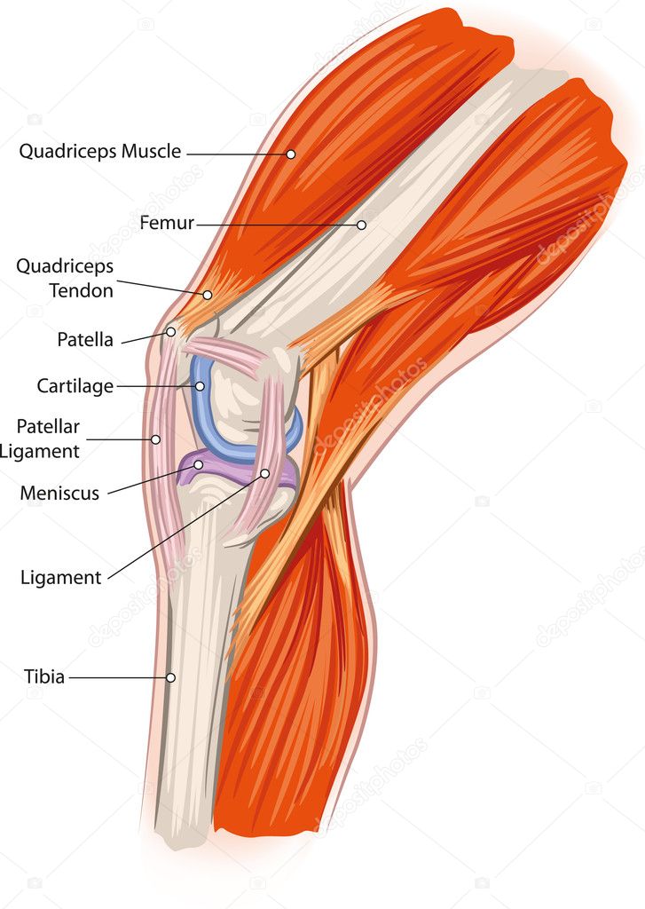
Knee anatomy Stock Vector Image by ©Lukaves 18341225
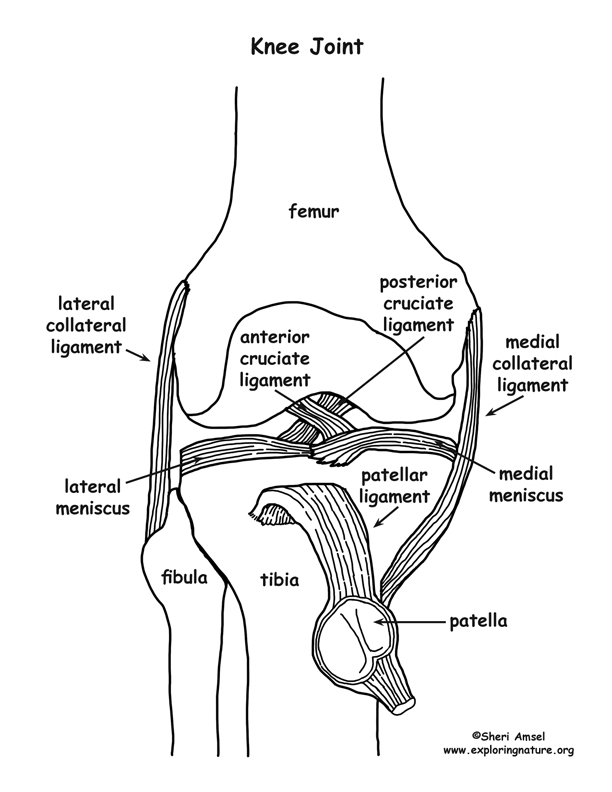
Knee Joint
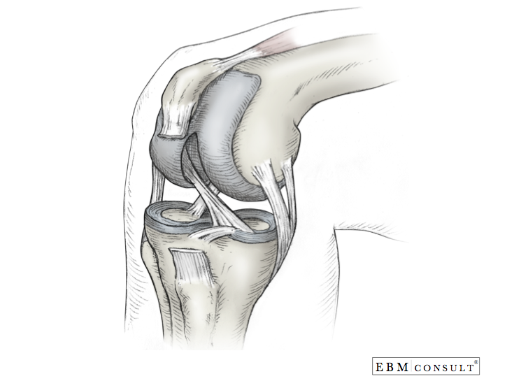
Anatomy Knee

FileKnee diagram.svg Wikipedia
Knee Joint from Lateral Surface ClipArt ETC

Knee injuries causes, types, symptoms, knee injuries prevention & treatment

Structure of the human knee Stock Vector Image by ©Silbervogel 72406683

Knee bones and joint sketch human anatomy Vector Image
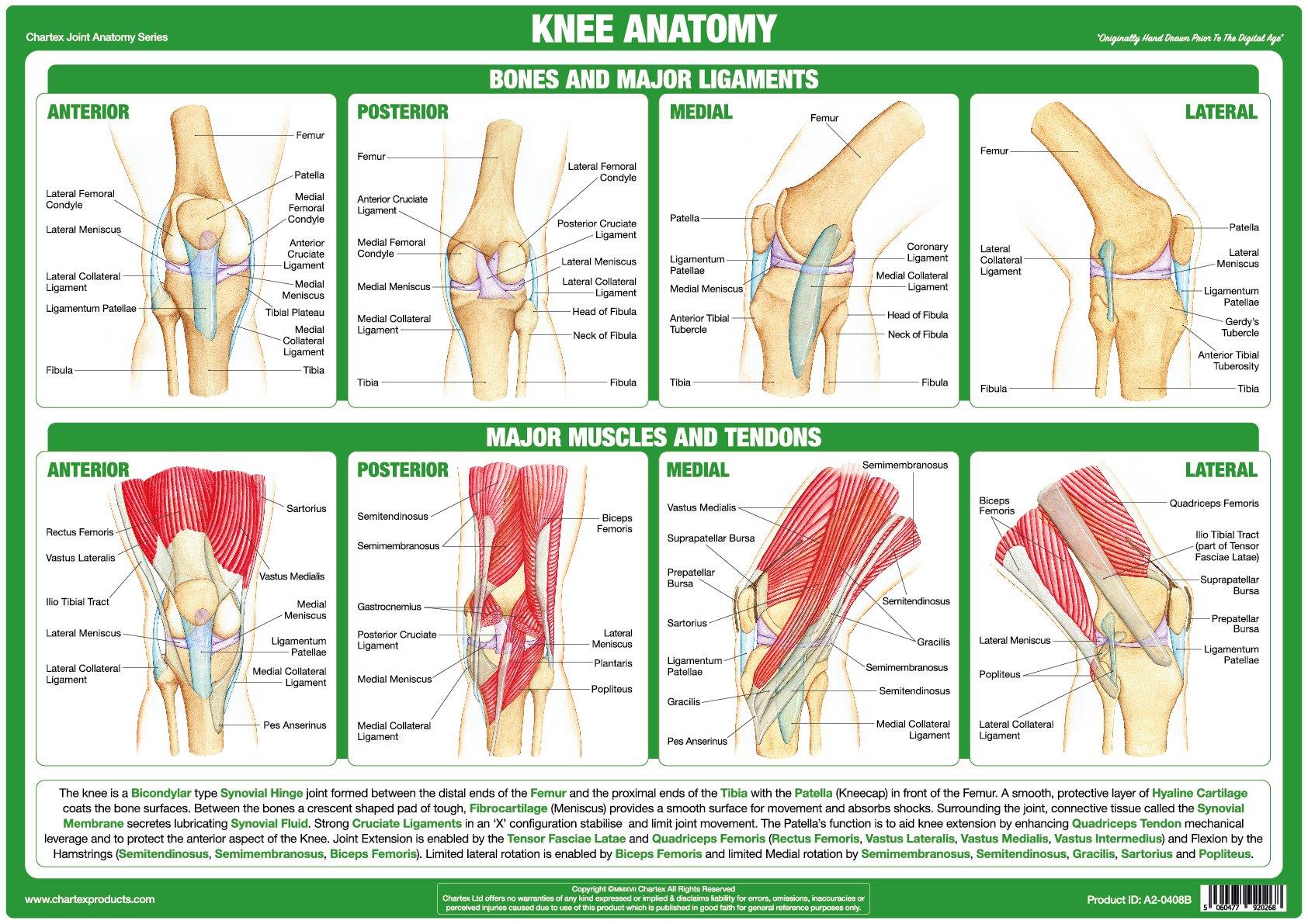
Knee Joint Anatomy Poster
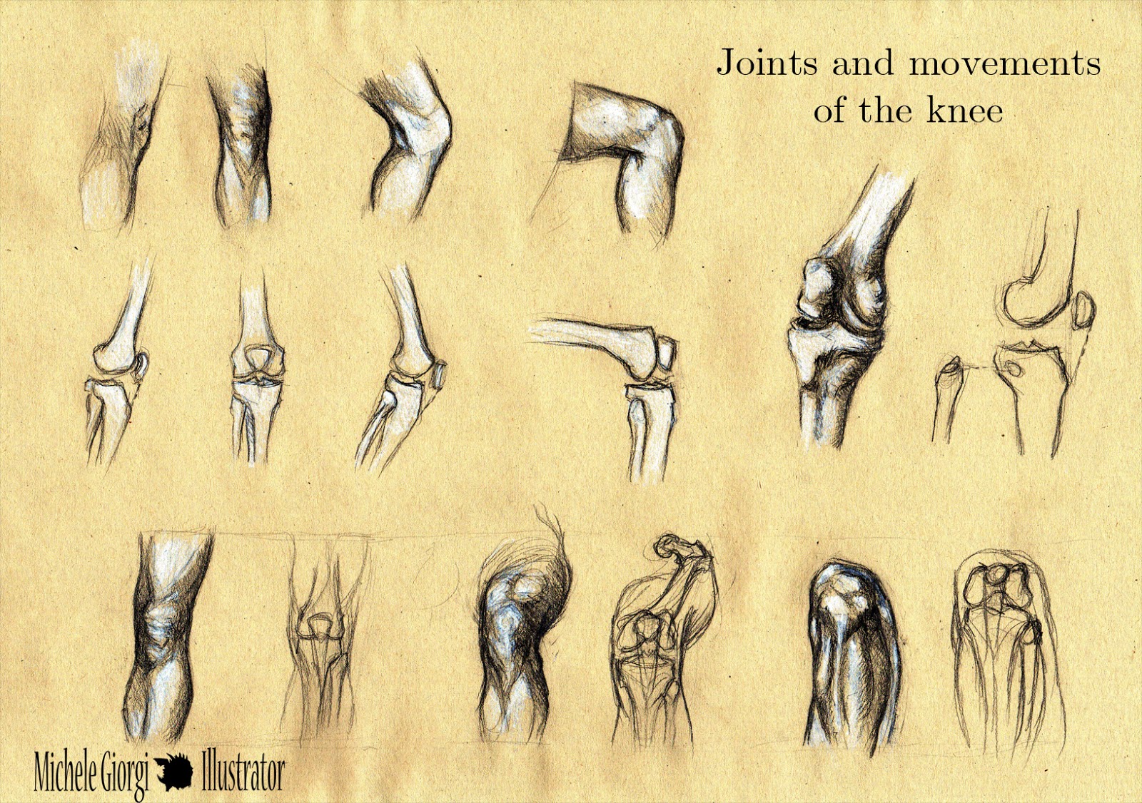
Michele Illustrator Anatomy Sketches Joints and movements of
Learn About The Muscles, Tendons, Bones, And Ligaments That Comprise The Knee Joint Anatomy.
Web The Anatomy Of The Knee Consists Of 3 Main Bones:
Web The Knee Joint Is A Hinge Type Synovial Joint, Which Mainly Allows For Flexion And Extension (And A Small Degree Of Medial And Lateral Rotation).
Two Rounded, Convex Processes (Known As Condyles) On The Distal End Of The Femur Meet Two Rounded, Concave Condyles At The Proximal End Of The Tibia.
Related Post: