Flow Chart For Gram Negative Bacteria
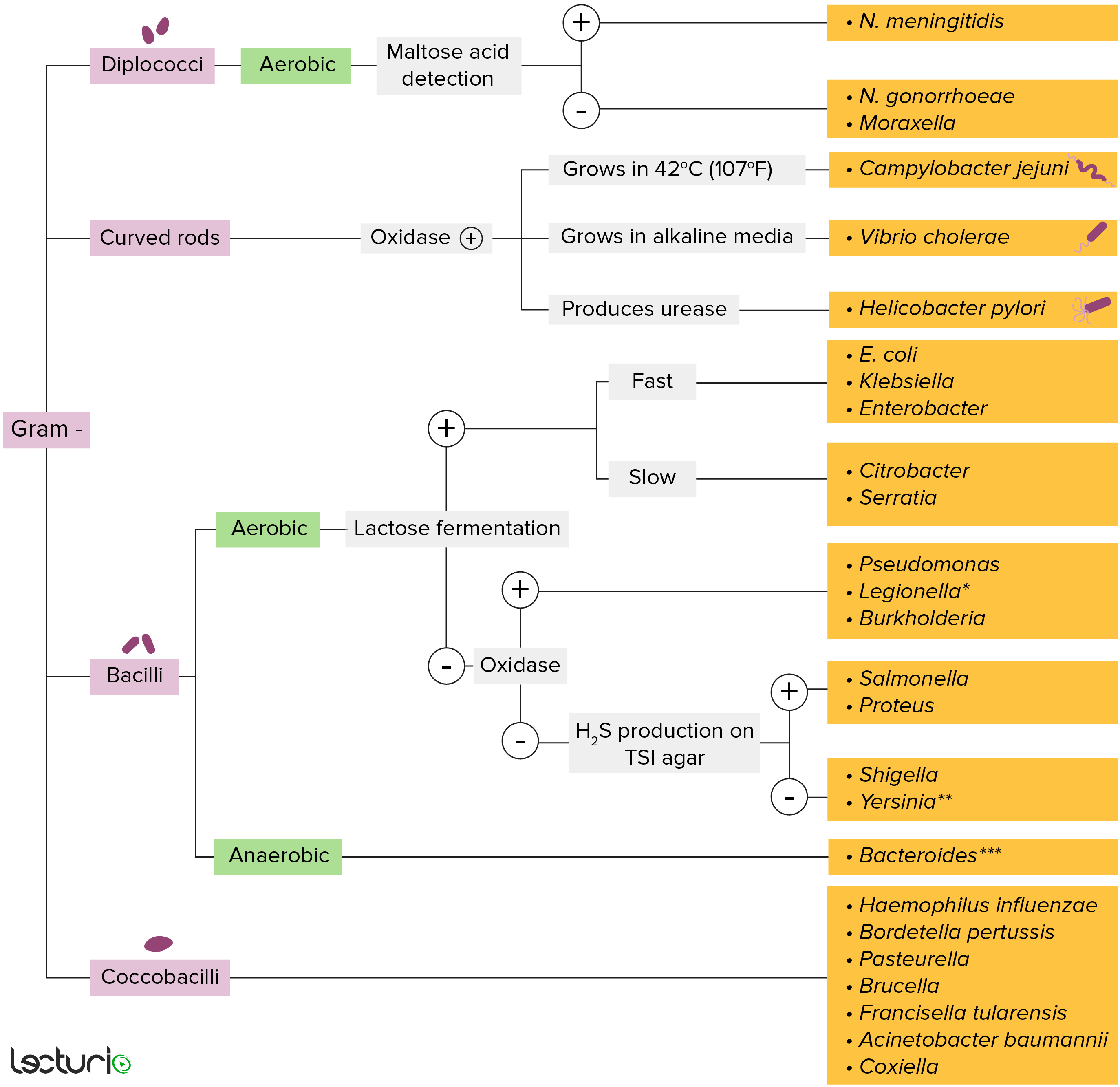
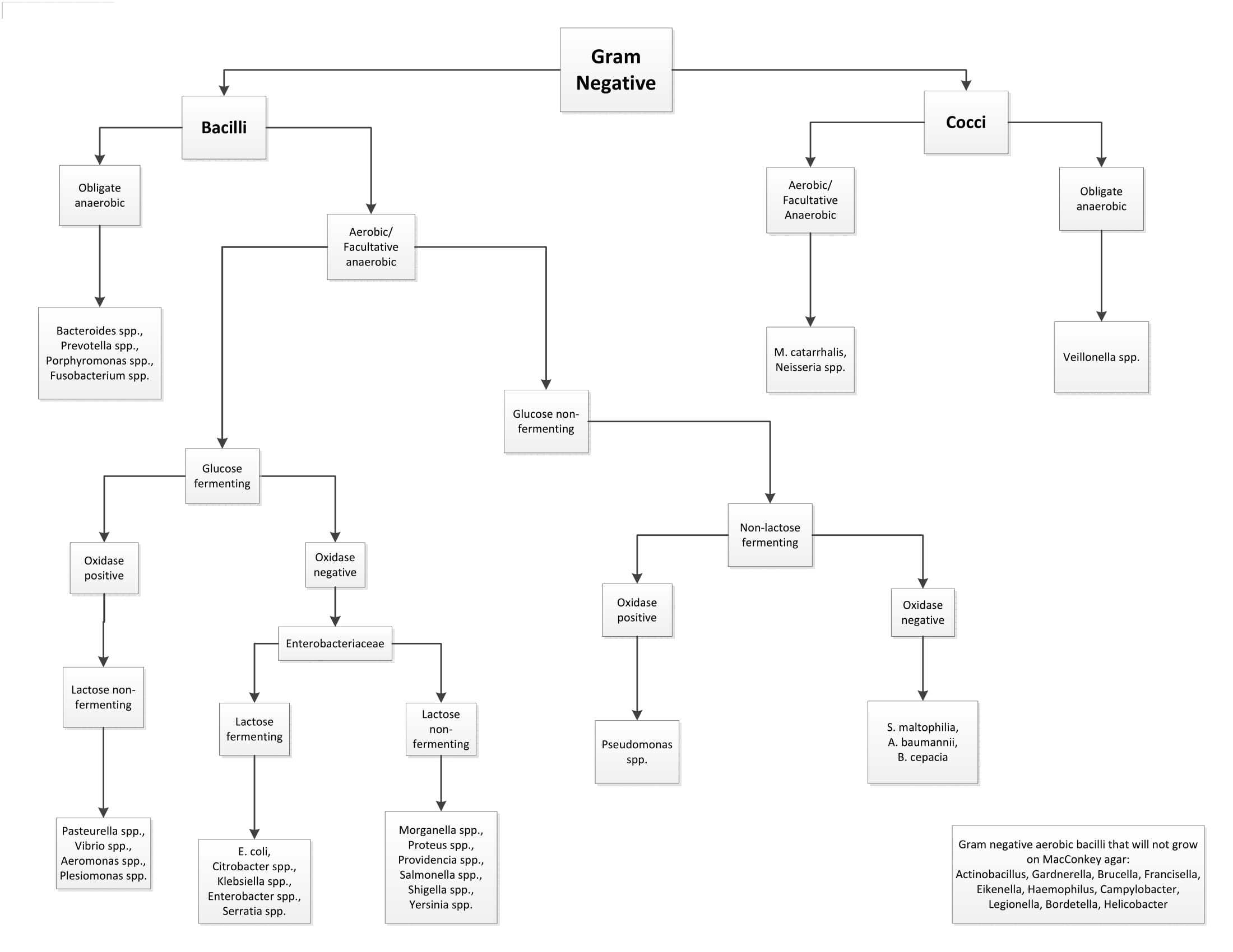
Flow Chart For Gram Negative Bacteria - Web gram stain lab report. (the graphic below is clickable. Gram staining is then used to identify if the bacteria are gram positive cocci, small gram positive bacilli, large gram positive bacilli, or gram negative bacilli. Move your mouse over an item on the graphic and if your arrow turns into a hand click on it and you will go to another place in the notebook.) click on gram negatives to determine how and when to perform the tests indicated above. The difference between the two groups is believed to be due to a much larger peptidoglycan (cell wall) in gram positives. Bacteria are identified in laboratories by various methods, including microscopy ( fresh state, after staining ), observation of growth characteristics ( list of culture media ), determination of reactions to organic and inorganic compounds ( api gallery , microbiological techniques) and molecular techniques. Use the results (positive or negative) from each test to determine the next test you should do on the flow chart. Web this new key provides a broad perspective of strategies useful for the biochemical identification of bacterial fish pathogens and it will be helpful in both diagnostic work and in teaching. The first test conducted was the gram stain test. Tmcc biology department microbiology resource center created date:. Know general characteristics of members of the enterobacteriaceae (e.g. This test differentiate the bacteria into gram positive and gram negative bacteria, which helps in the classification and differentiations of microorganisms. (the graphic below is clickable. Web start studying gram negative bacteria flow chart. The difference between the two groups is believed to be due to a much larger peptidoglycan (cell. Know general characteristics of members of the enterobacteriaceae (e.g. Colonial morphology and oxidase reactivity determine the pathway to be followed on the flow chart which then indicates the specific test to be perform. Web gram negative flow chart. Identify different types of colonial characteristics. Web gram negative aerobic bacilli that will not grow on macconkey agar: Gram project is a medical education resource website containing diagrams, tables and flowcharts for all your quick referencing, revision and teaching needs. Materials based off of the flow chart and the methods took to identify the two unknowns the tests i conducted were the gram stain test, endospore stain test, urease test, lactase test, and lastly the mrvp methyl red. * = see biochemical tests for gram negative organism id job aid for positive and negative result reference. This capsule helps prevent white blood cells (which fight infection) from ingesting the bacteria. Web gram staining is the common, important, and most used differential staining techniques in microbiology, which was introduced by danish bacteriologist hans christian gram in 1884. Web gram. Learn vocabulary, terms, and more with flashcards, games, and other study tools. Materials based off of the flow chart and the methods took to identify the two unknowns the tests i conducted were the gram stain test, endospore stain test, urease test, lactase test, and lastly the mrvp methyl red test. Web gram negative flow chart. Web gram negative aerobic. Materials based off of the flow chart and the methods took to identify the two unknowns the tests i conducted were the gram stain test, endospore stain test, urease test, lactase test, and lastly the mrvp methyl red test. Tmcc microbiology resource center unknown identification work flow flowchart. Web gram negative bacteria types and classification flowchart. Web gram negative rods. This capsule helps prevent white blood cells (which fight infection) from ingesting the bacteria. Gram project is a medical education resource website containing diagrams, tables and flowcharts for all your quick referencing, revision and teaching needs. Web gram negative bacteria types and classification flowchart. Web the flow chart of the procedure is shown below. Use the results (positive or negative). * = see biochemical tests for gram negative organism id job aid for positive and negative result reference. Gram staining is then used to identify if the bacteria are gram positive cocci, small gram positive bacilli, large gram positive bacilli, or gram negative bacilli. Web gram negative bacteria types and classification flowchart. Price chart facebook twitter tiktok help faq platform. Gram neg., oxidase negative, fermentative, opportunistic and primary infections). Unknown identification work flow flowchart author: Gram project is a medical education resource website containing diagrams, tables and flowcharts for all your quick referencing, revision and teaching needs. Web gram negative flow chart. Samples are placed on mac conkey agar and blood agar to observe growth and colony appearance. Use flowcharts and identification charts to identify some common aerobic gram negative microorganisms. This test differentiate the bacteria into gram positive and gram negative bacteria, which helps in the classification and differentiations of microorganisms. Know general characteristics of members of the enterobacteriaceae (e.g. * = see biochemical tests for gram negative organism id job aid for positive and negative result. Web gram negative flow chart. Web gram negative rods stool pathogens flowchart. Actinobacillus, gardnerella, brucella, francisella, eikenella, haemophilus, campylobacter, legionella, bordetella, helicobacter. 1 = note the odor without sniffing the plate. Gram staining is then used to identify if the bacteria are gram positive cocci, small gram positive bacilli, large gram positive bacilli, or gram negative bacilli. Web this flow chart outlines the steps to identify bacteria from a specimen. Identify different types of bacterial morphology seen on a gram stain. Bacteria are identified in laboratories by various methods, including microscopy ( fresh state, after staining ), observation of growth characteristics ( list of culture media ), determination of reactions to organic and inorganic compounds ( api gallery , microbiological techniques) and molecular techniques. Web gram negative aerobic bacilli that will not grow on macconkey agar: Web at the conclusion of this elearning, you should be able to: Gram project is a medical education resource website containing diagrams, tables and flowcharts for all your quick referencing, revision and teaching needs. Know the basis of the serological classification. Gram neg., oxidase negative, fermentative, opportunistic and primary infections). Samples are placed on mac conkey agar and blood agar to observe growth and colony appearance. Move your mouse over an item on the graphic and if your arrow turns into a hand click on it and you will go to another place in the notebook.) click on gram negatives to determine how and when to perform the tests indicated above. The first test conducted was the gram stain test.
Gram negative rods flowchart labquiz

Biochemical Gram Negative Chart

Top Unknown Gram Negative Bacteria Flow Chart Wallpapers Medical

Gram neg flow chart Med tech Pinterest

Bacteroides Concise Medical Knowledge

Gram Negative Flow Chart 2 Diagram Quizlet

Gram Negative Bacteria Flow Chart

Pin by Rachel Noble on MICROBIOLOGY rotation Pinterest

19 Awesome Gram Negative Flow Chart Labb by AG

Gram Negative Rods Flow Chart vrogue.co
Unknown Identification Work Flow Flowchart Author:
Web Gram Stain Lab Report.
Web The Flow Chart Of The Procedure Is Shown Below.
Tmcc Biology Department Microbiology Resource Center Created Date:.
Related Post: