Eye Anatomical Chart
Eye Anatomical Chart - The eye anatomical chart illustrates anterior chamber angle, lens, retina, fundus and the macula lutea. Clear and flexible, the lens changes shape to focus light on the retina. The anatomy of the eye includes auxiliary structures, such as the bony eye socket and extraocular muscles, as well as the structures of the eye itself, such as the lens and the retina. Read an article about how vision works. Web 6 min read. Web at hoffner eye care we strive to give you an unparalleled experience from routine eye exams, contact lens fittings, treatment and management of ocular diseases; Web this popular chart of the eye has illustrations by award winning medical illustrator keith kasnot. Whether you are an optometrist or professor of human anatomy, use our detailed standard eye charts for sale or 3d model of the eye for classroom teaching or patient education. Web interactive ophthalmic figures for medical student education illustrate concepts in eye anatomy and functions in an engaging format. Web the eye anatomical chart shows cross section of the eye. Web human eye, specialized sense organ in humans that is capable of receiving visual images, which are relayed to the brain. The eye is cushioned within the orbit by pads of fat. A diagram to learn about the parts of the eye and what they do. Here is a tour of the eye starting from the outside, going in through. This chart focusses on the anatomy of the eye as it relates to the most frequent causes of eye diseases and is targeted at medical students, trainee ophthalmologists, optometrists, and other eye care specialists. The chart also provides lateral and top view of the eye and shows the visual field. The medical information on this site is provided as an. The medical information on this site is provided as an information resource only, and is not to. In addition to the eyeball itself, the orbit contains the muscles that move the eye, blood vessels, and nerves. Web click on various parts of our human eye illustration for descriptions of the eye anatomy; The eye anatomical chart illustrates anterior chamber angle,. Illustrates anterior chamber angle, lens, retina, fundus and the macula lutea. The front part (what you see in the mirror) includes: Your eye is a slightly asymmetrical globe, about an inch in diameter. Web 6 min read. Web learn about the anatomy of the human eye from the specialists at florida eye clinic, serving patients in central florida. The front part (what you see in the mirror) includes: In addition to the eyeball itself, the orbit contains the muscles that move the eye, blood vessels, and nerves. Only the most important anatomical details are listed, alongside web links to videos and diagrams (each underlined text is a link) Your eye is a slightly asymmetrical globe, about an inch. Shows cross section of the eye. Web to understand the diseases and conditions that can affect the eye, it helps to understand basic eye anatomy. Web human eye, specialized sense organ in humans that is capable of receiving visual images, which are relayed to the brain. In addition to the eyeball itself, the orbit contains the muscles that move the. Junction of the front surface of the iris and the back surface of the cornea, where aqueous fluid filters out of the eye. Read an overview of general eye anatomy to learn how the parts of the eye work together. Read an article about how vision works. The conjunctiva is the membrane covering the sclera (white portion of your eye).. A clear dome over the iris. Carries the messages from the retina to the brain. Web below, find an explanation of each anatomical part from the above video with physiologic and pathologic correlates. Web 6 min read. Also provides lateral and top view of the eye and shows the visual field. A diagram to learn about the parts of the eye and what they do. Web learn about the anatomy of the human eye from the specialists at florida eye clinic, serving patients in central florida. Eyes are approximately one inch in diameter. Also provides lateral and top view of the eye and shows the visual field. Illustrates anterior chamber angle,. This chart focusses on the anatomy of the eye as it relates to the most frequent causes of eye diseases and is targeted at medical students, trainee ophthalmologists, optometrists, and other eye care specialists. Web click on various parts of our human eye illustration for descriptions of the eye anatomy; Web the structures of the eye include the cornea, iris,. Pads of fat and the surrounding bones of the. Web to understand the diseases and conditions that can affect the eye, it helps to understand basic eye anatomy. Carries the messages from the retina to the brain. To the choice of the correct frame and lenses according to your prescription and lifestyle. Here is a tour of the eye starting from the outside, going in through the front and working to the back. Also provides lateral and top view of the eye and shows the visual field. Junction of the front surface of the iris and the back surface of the cornea, where aqueous fluid filters out of the eye. Part of the teachme series. A diagram to learn about the parts of the eye and what they do. Read an overview of general eye anatomy to learn how the parts of the eye work together. Illustrates cataract, corneal ulcers, retinal tear and detachment, vitreous floaters, glaucoma, melanoma, diabetic. Web interactive ophthalmic figures for medical student education illustrate concepts in eye anatomy and functions in an engaging format. Read an article about how vision works. Web at hoffner eye care we strive to give you an unparalleled experience from routine eye exams, contact lens fittings, treatment and management of ocular diseases; Web the eye has several major components: Web the eye anatomical chart.
Human Eye Chart at Rs 800/piece Human Eye Chart in Ambala ID
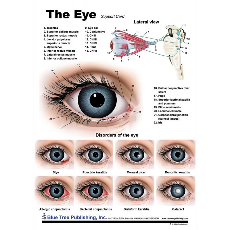
Eye Anatomical Chart
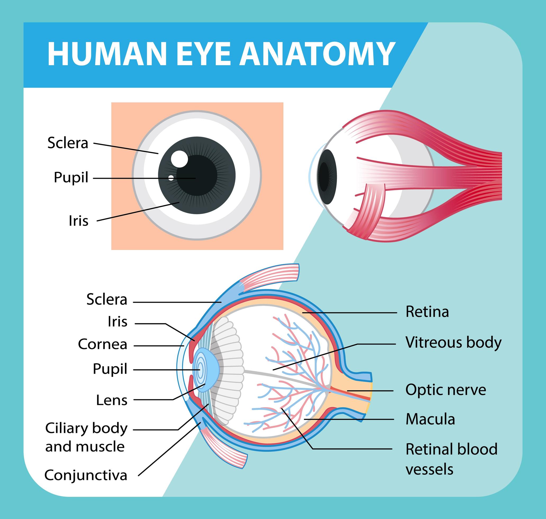
Diagram of human eye anatomy with label 1848847 Vector Art at Vecteezy

Eye Anatomical Poster Clinical Charts and Supplies
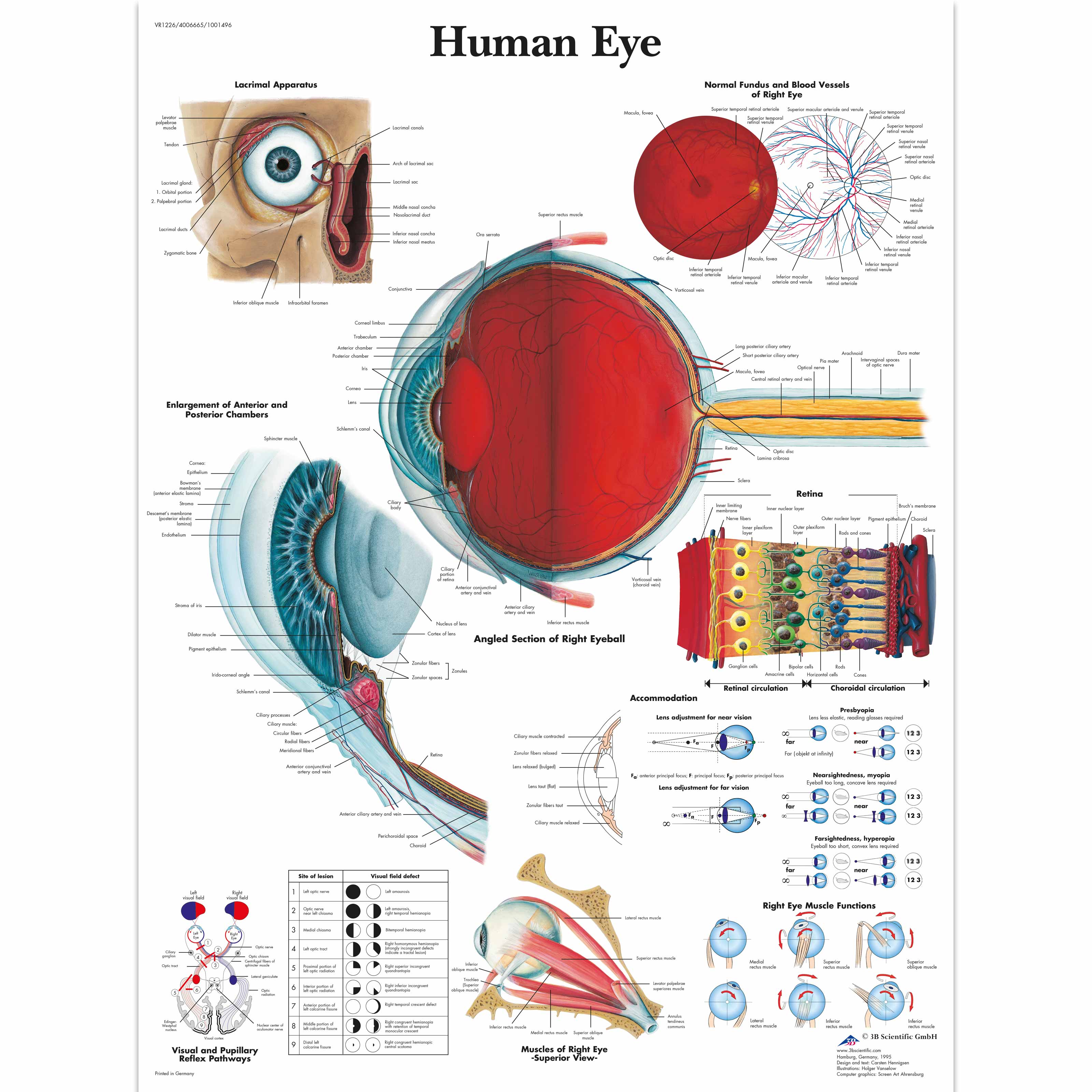
Human Eye Chart 1001496 3B Scientific VR1226L Ophthalmology

Scientific Publishing The Eye Anatomy Chart

Eye Anatomical Chart
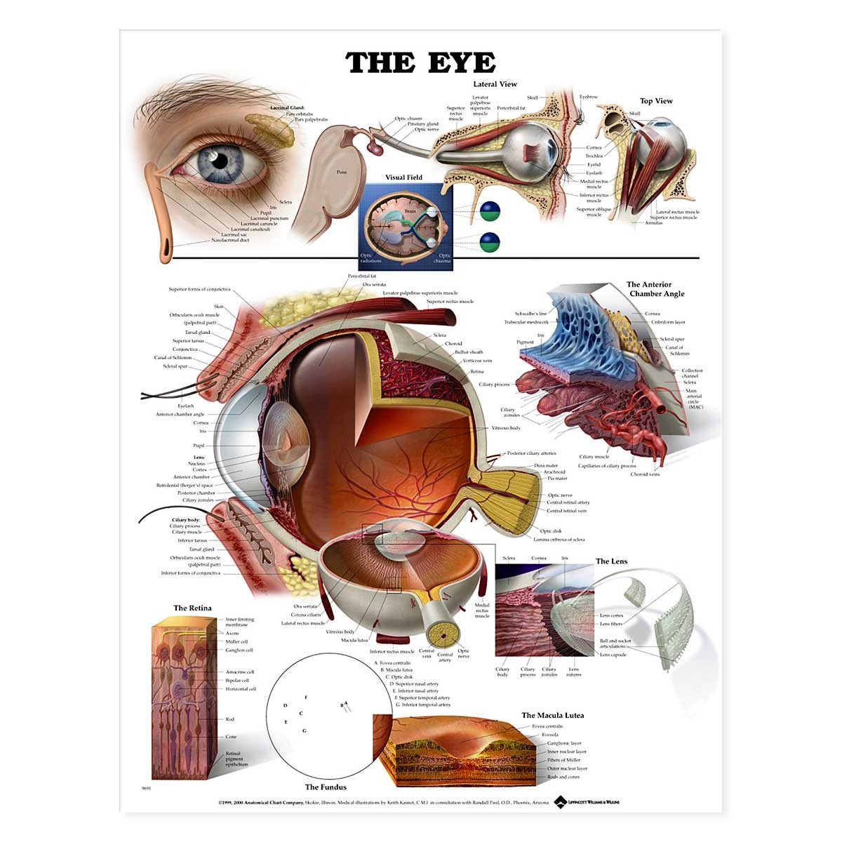
The Eye Anatomical Chart 20'' x 26''

Eye Anatomy & Pathology Collection Pupil Cataract Models Charts
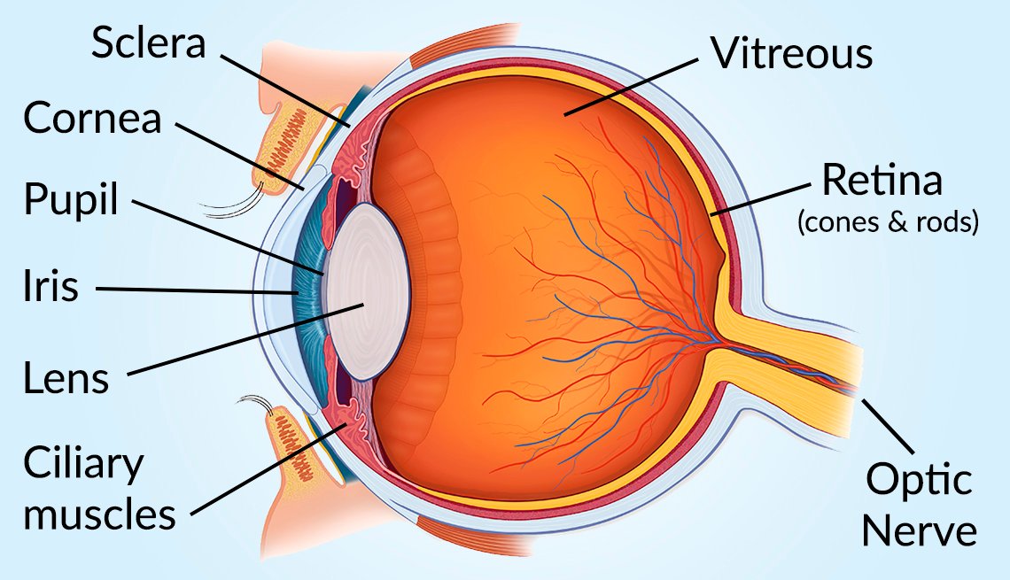
Vision and Eye Diagram How We See
Shows Cross Section Of The Eye.
The Chart Also Provides Lateral And Top View Of The Eye And Shows The Visual Field.
Web This Popular Chart Of The Eye Has Illustrations By Award Winning Medical Illustrator Keith Kasnot.
Whether You Are An Optometrist Or Professor Of Human Anatomy, Use Our Detailed Standard Eye Charts For Sale Or 3D Model Of The Eye For Classroom Teaching Or Patient Education.
Related Post: