Ear Chart
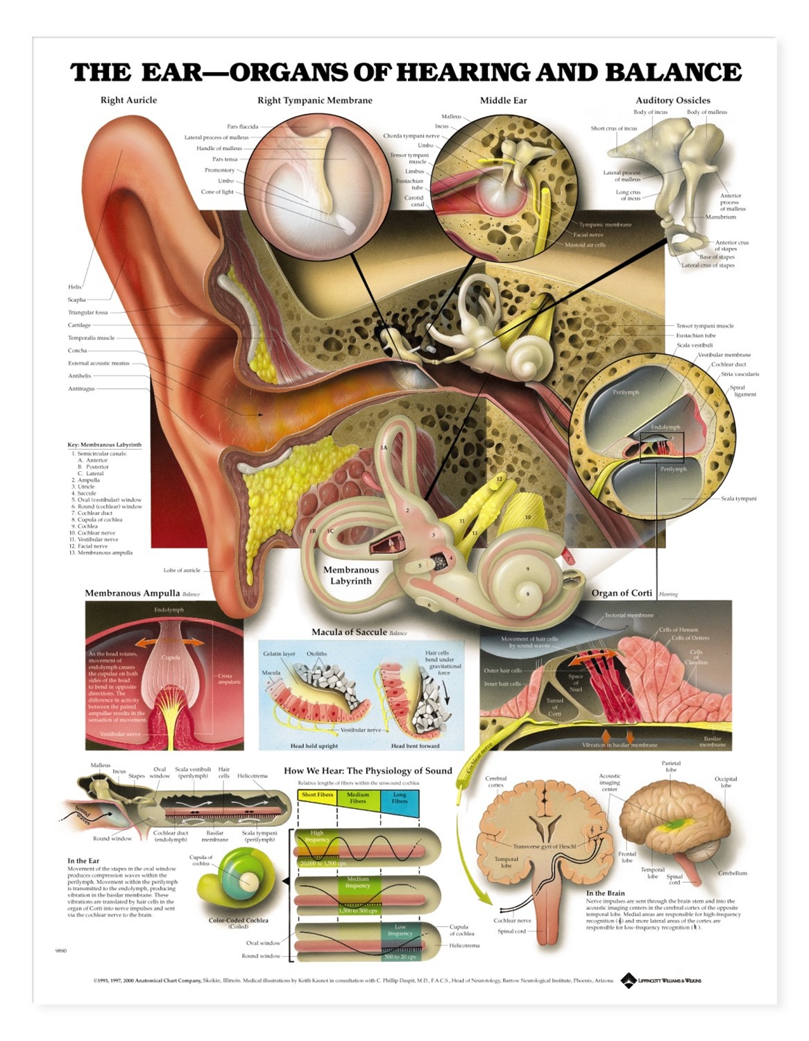
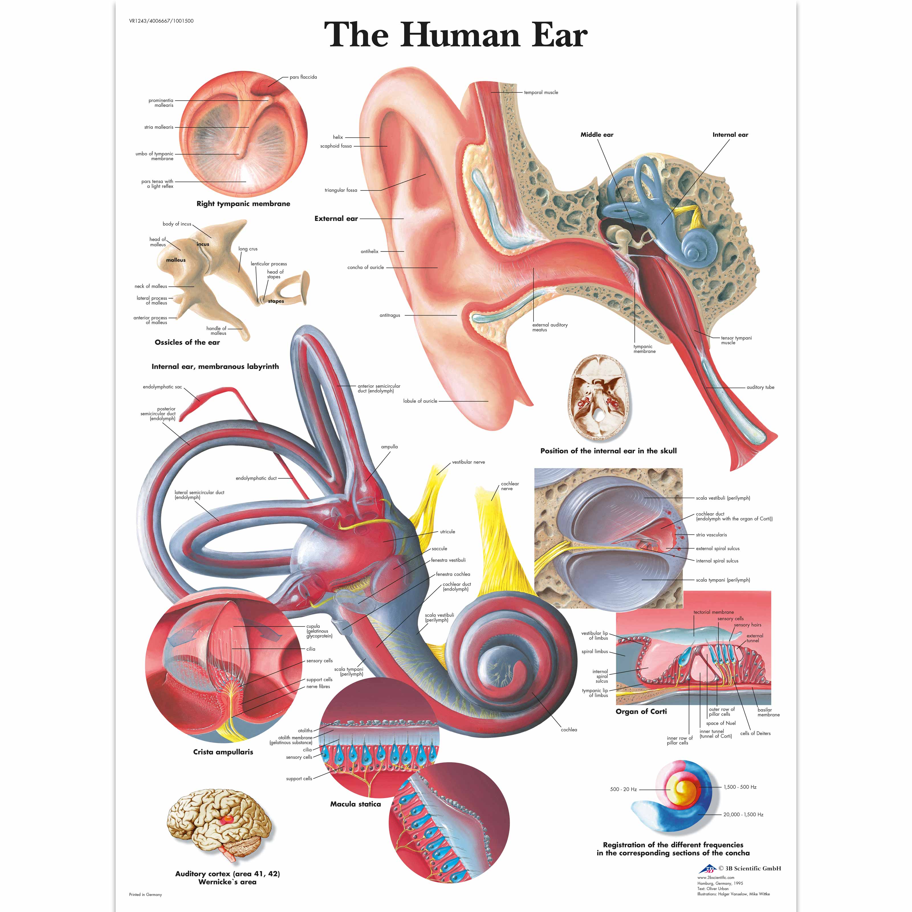
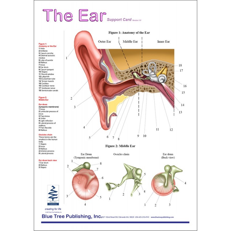
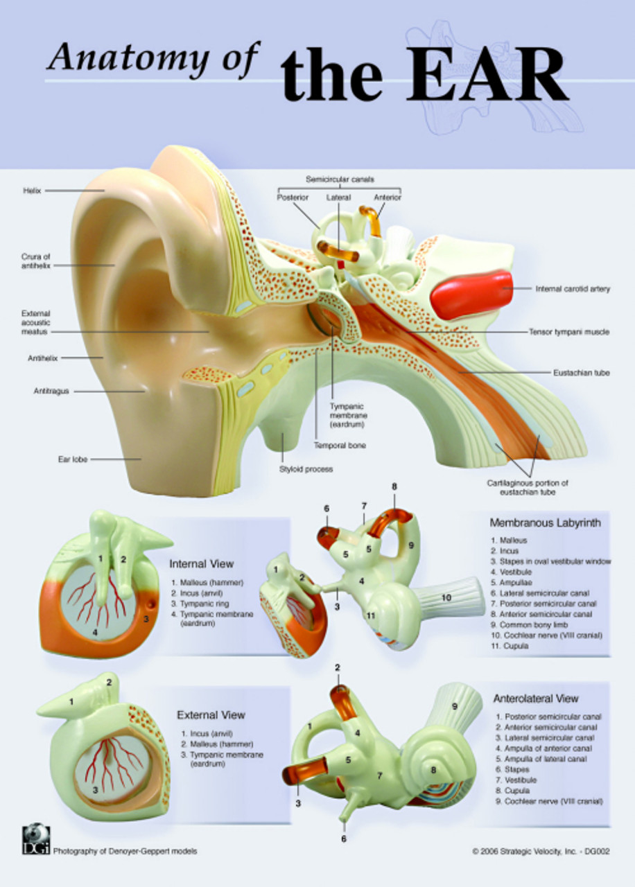
Ear Chart - The external ear can be divided functionally and structurally into two parts; It comprises a pinna, external auditory meatus (canal) & tympanic membrane. The ear consists of external, middle, and inner structures. This structure helps to give each of us our unique appearance. Web the ear organs of hearing and balance anatomical chart illustrates ear anatomy including right auricle, right tympanic membrane, middle ear, auditory ossicles, membranous labryinth, membranous ampulla, organ of corti, macula of saccule. The outer ear comes in all types of shapes and sizes. Look for an l or r at the top right to identify the eardrum tested. Web the ear canal, or auditory canal, is a tube that runs from the outer ear to the eardrum. The eardrum vibrates when sound waves enter. Web the ear is the sensory organ for hearing and balance and it is anatomically divided into 3 parts: The outer ear comes in all types of shapes and sizes. Learn about the anatomy and physiology of the human ear in this article. Medically reviewed by jennifer boidy, rn. Web the three main parts of your ear include the outer ear, middle ear and inner ear. 3 performing an ear reflexology massage. Your tympanic membrane (eardrum) separates your outer ear and middle ear. Web fbi director christopher wray revealed the gunman who shot donald trump did research on john kennedy's assassin and flew a drone over the rally area. The ears are organs that provide two main functions — hearing and balance — that depend on specialized receptors called hair cells. The. Outer ear (external ear) your outer ear is the part of your ear that’s visible. Web human ear, organ of hearing and equilibrium that detects and analyzes sound by transduction and maintains the sense of balance. This structure helps to give each of us our unique appearance. Web each ear consists of three portions: Web an overview of the anatomy. The medical term for the outer ear is the auricle or pinna. The pinna is a projecting elastic cartilage covered with skin. Its most prominent outer ridge is called the helix. The outer, middle, and inner ear. The external ear can be divided functionally and structurally into two parts; Web the three main parts of your ear include the outer ear, middle ear and inner ear. The outwardly visible part of the ear is composed of skin and cartilage, and attaches to the skull. The poster presents information about the human ear in colorful detail. Web human ear chart | ear, nose and throat (ent) | this anatomical chart. It comprises a pinna, external auditory meatus (canal) & tympanic membrane. The outer, middle, and inner ear. L indicates results for the left eardrum and r indicates results for the right eardrum. The outer ear comes in all types of shapes and sizes. The medical term for the outer ear is the auricle or pinna. The external, middle and internal ear. How to protect your ears. Paul nogier's ear map was first published in 1957. Outer ear (external ear) your outer ear is the part of your ear that’s visible. The poster presents information about the human ear in colorful detail. Web the three main parts of your ear include the outer ear, middle ear and inner ear. Web paul nogier, md, discovered ear somatotopy, a representation of the whole person on the ear in the shape of a homunculus, or inverted fetus, on the ear. At the evening rally, trump remained largely tied to his prepared remarks and attacked harris. This structure helps to give each of us our unique appearance. The eardrum vibrates when sound waves enter. The ear canal and outer cartilage of the ear make up the. The outer ear comes in all types of shapes and sizes. The medical term for the outer ear is the auricle or pinna. Web the ear organs of hearing and balance anatomical chart illustrates ear anatomy including right auricle, right tympanic membrane, middle ear, auditory ossicles, membranous labryinth, membranous ampulla, organ of corti, macula of saccule. Learn about the anatomy and physiology of the human ear in this article. Web the three main parts of your ear include the outer ear, middle ear. Web paul nogier, md, discovered ear somatotopy, a representation of the whole person on the ear in the shape of a homunculus, or inverted fetus, on the ear. It’s what most people mean when they say “ear.” Locate the vertical y axis to find the compliance of the eardrum. 2 reading an emotional ear reflexology chart. The ear is divided into three portions: Web discover how, why, where and when hearing loss can occur within the ear. The outer ear, the middle ear, and the inner ear. The outer ear is made up of cartilage and skin. Web human ear, organ of hearing and equilibrium that detects and analyzes sound by transduction and maintains the sense of balance. Outer ear (external ear) your outer ear is the part of your ear that’s visible. The outer, middle, and inner ear. The external ear can be divided functionally and structurally into two parts; Your tympanic membrane (eardrum) separates your outer ear and middle ear. Paul nogier's ear map was first published in 1957. The medical term for the outer ear is the auricle or pinna. Medically reviewed by jennifer boidy, rn.
The Ear Organs of Hearing and Balance Anatomical Chart Physio Needs

Anatomical Charts and Posters Anatomy Charts The Human Ear Chart

Ear Reflexology Chart Accurate Description Corresponding Stock Vector

Ear Anatomical Chart

Chart with Reflexology 🥇 Ear Preassure Points 【2020

Buy Ultimate Auriculotherapy Reference Card, Showing All Ear Points

Hearing System Chart Anatomy of the Ear Etsy in 2022 Anatomy

Ear Anatomy Chart

Anatomy Of the Ear Notebook Size Charts Clinical Charts and Supplies

Human Ear Chart ubicaciondepersonas.cdmx.gob.mx
The Pinna Is A Projecting Elastic Cartilage Covered With Skin.
L Indicates Results For The Left Eardrum And R Indicates Results For The Right Eardrum.
Web The Ear Organs Of Hearing And Balance Anatomical Chart Illustrates Ear Anatomy Including Right Auricle, Right Tympanic Membrane, Middle Ear, Auditory Ossicles, Membranous Labryinth, Membranous Ampulla, Organ Of Corti, Macula Of Saccule.
The Inner Ear (Aka Labyrinth) Is The Deepest Part Of The Ear And Plays An Essential Role In Hearing And Balance.
Related Post: