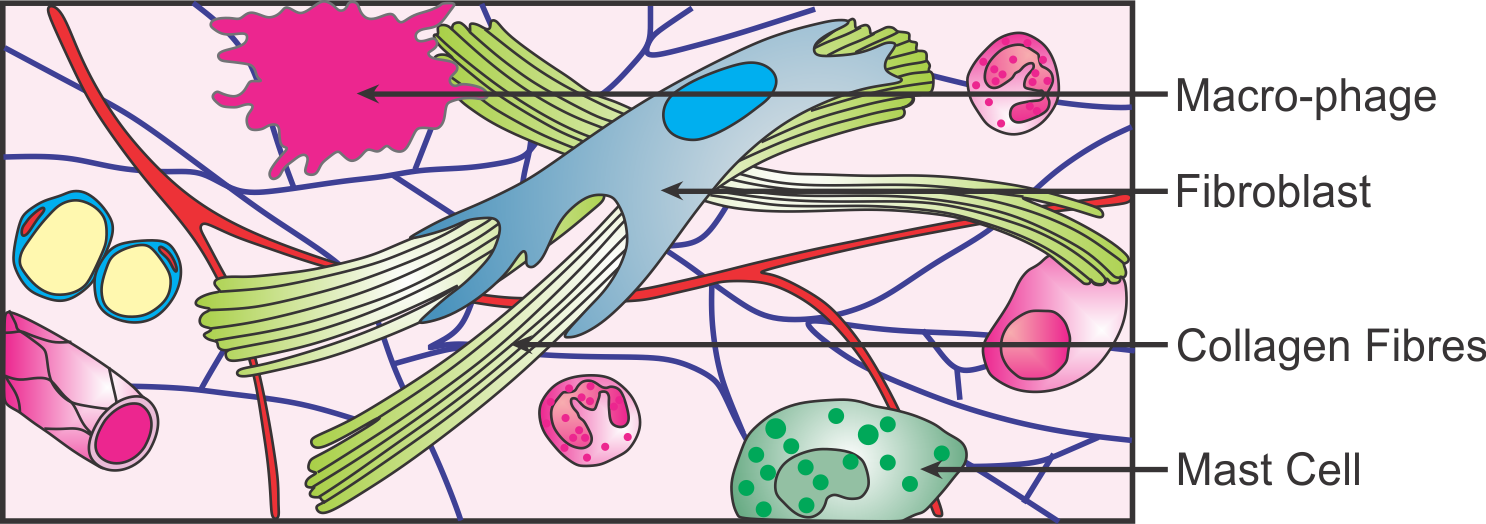
Areolar Connective Tissue Drawing
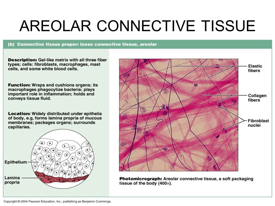
Areolar Connective Tissue Drawing - Its ground substance occupies more volume than the fibers do. They usually stain pink and are the thickest fiber. Web #biology #class9 #class11 #areolarconnectivetissue #animaltissues #hscbiology #maharashtrastateboard2021 #biology2021 #biologydiagrams #icse #cbsethis video. Macrophages are present as well. It is the most widely encountered type of connective tissue and contains most of the connective tissue components. Cells, protein fibers, and an amorphous ground substance. Web how to draw a diagram of areolar tissue in exam is the topic. Together the fibers and ground substance make up the extracellular matrix. Connective tissuehistology drawing eith explanation loose areolar tissueif you found helpfulhistology drawing of loose areolar tissue with expla. Web recognize different types of connective tissue (e.g., dense irregular, dense regular, loose, adipose) and know examples where they are found in the body. Web loose connective tissue (lct), also called areolar tissue, belongs to the category of connective tissue proper. These serve to hold organs and other tissues in place and, in the case of adipose tissue, isolate and store energy reserves. Connective tissuehistology drawing eith explanation loose areolar tissueif you found helpfulhistology drawing of loose areolar tissue with expla. The important function. Web after the epithelium, i am posting videos on how to draw connective tissue. The important function of this type of tissue is that it provides nutrition to the cells and also acts as a cushion to protect the. Recognize basement membranes (or basal lamina) in light micrograph and em sections and know their functions. Web loose connective tissue, also. Web the papillary layer is made of loose, areolar connective tissue, which means the collagen and elastin fibers of this layer form a loose mesh. Web after the epithelium, i am posting videos on how to draw connective tissue. Web loose connective tissue, also known as areolar tissue, is a cellular connective tissue with thin and relatively sparse collagen fibers.. This loose fibrous tissue is widely distributed in the body where it provides strength, elasticity, and support to neighboring tissues. They usually stain pink and are the thickest fiber. This is the well labelled diagram of structure of areolar tissue. The important function of this type of tissue is that it provides nutrition to the cells and also acts as. Loose connective tissue has some fibroblasts, although macrophages are present as well. Web study with quizlet and memorize flashcards containing terms like fibroblast, macrophages, mast cells and more. As illustrated in figure 1, loose connective tissue has some fibroblasts; This loose fibrous tissue is widely distributed in the body where it provides strength, elasticity, and support to neighboring tissues. They. Web let's explore what areolar connective tissue looks like and what it does. The ecm is composed of a moderate amount of ground substance and two main types of protein fibers: These serve to hold organs and other tissues in place and, in the case of adipose tissue, isolate and store energy reserves. Its cellular content is highly abundant and. Recognize basement membranes (or basal lamina) in light micrograph and em sections and know their functions. It is the most widely encountered type of connective tissue and contains most of the connective tissue components. Macrophages are present as well. Its ground substance occupies more volume than the fibers do. Web areolar tissue is the least specialized type of connective tissue. Web loose connective tissue, also called areolar connective tissue, has a sampling of all of the components of a connective tissue. Macrophages are present as well. Collagen fibers are found in most supporting tissues and collagen is the most abundant protein in the body (wheaters). Web loose connective tissue, also called areolar connective tissue, has a sampling of all of. Connective tissuehistology drawing eith explanation loose areolar tissueif you found helpfulhistology drawing of loose areolar tissue with expla. Web #biology #class9 #class11 #areolarconnectivetissue #animaltissues #hscbiology #maharashtrastateboard2021 #biology2021 #biologydiagrams #icse #cbsethis video. The ecm is composed of a moderate amount of ground substance and two main types of protein fibers: Cells, protein fibers, and an amorphous ground substance. This loose fibrous. Web study with quizlet and memorize flashcards containing terms like fibroblast, macrophages, mast cells and more. Web loose connective tissue, also called areolar connective tissue, has a sampling of all of the components of a connective tissue. Web photographs of loose, areolar connective tissue in the intestine (lamina propria), skin (adipose, hypodermis) and spleen (reticular fibers) Connective tissuehistology drawing eith. Loose connective tissue has some fibroblasts, although macrophages are present as well. Web #biology #class9 #class11 #areolarconnectivetissue #animaltissues #hscbiology #maharashtrastateboard2021 #biology2021 #biologydiagrams #icse #cbsethis video. Collagen fibers are found in most supporting tissues and collagen is the most abundant protein in the body (wheaters). Cells, protein fibers, and an amorphous ground substance. As illustrated in figure 1, loose connective tissue has some fibroblasts; Web after the epithelium, i am posting videos on how to draw connective tissue. As illustrated in figure 1, loose connective tissue has some fibroblasts; This is the well labelled diagram of structure of areolar tissue. This superficial layer of the dermis projects into the stratum basale of the epidermis to. They usually stain pink and are the thickest fiber. Web loose connective tissue, also called areolar connective tissue, has a sampling of all of the components of a connective tissue. These serve to hold organs and other tissues in place and, in the case of adipose tissue, isolate and store energy reserves. Recognize basement membranes (or basal lamina) in light micrograph and em sections and know their functions. Web the term areolar connective tissue means tissue with 'small open spaces' (areola) and refers to the appearance of small airy pockets between the network of cells and fibers. Web areolar tissue, found in the hypodermis of the skin and below the epithelial layers of the digestive, respiratory, and urinary tracts, is a loose connective tissue proper, as is adipose tissue, also known as fat. Web loose connective tissue, also known as areolar tissue, is a cellular connective tissue with thin and relatively sparse collagen fibers.
With Help Of Neat Labelled Diagram Describe The Struc vrogue.co
Areolar Connective Tissue

Areolar Connective Tissue Diagram

Areolar Connective Tissue Diagram

chapter 4 connective tissues neuron stuff and other science stuff


Histology Drawing of Loose Areolar Tissue with explanation connective

How to draw areolar tissue most easy way YouTube

Function of Areolar Connective Tissue Video & Lesson Transcript

Areolar Connective Tissue Labeled Diagram
Web Loose Connective Tissue (Lct), Also Called Areolar Tissue, Belongs To The Category Of Connective Tissue Proper.
Web Loose Connective Tissue, Also Called Areolar Connective Tissue, Has A Sampling Of All Of The Components Of A Connective Tissue.
Together The Fibers And Ground Substance Make Up The Extracellular Matrix.
Macrophages Are Present As Well.
Related Post:
