Anatomy Of Heart Drawing
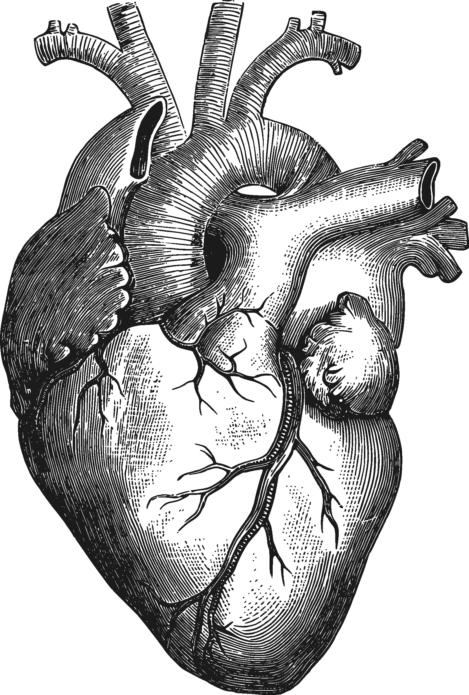
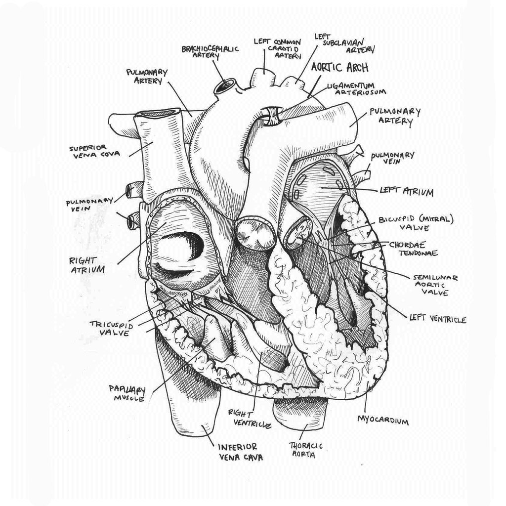
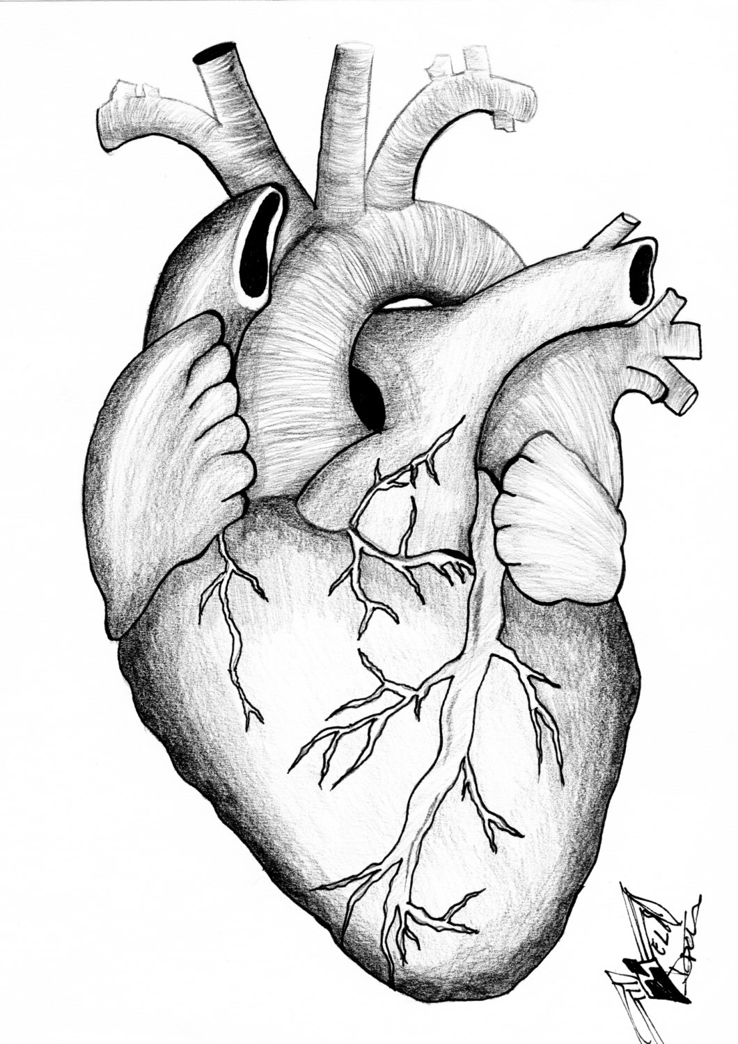
Anatomy Of Heart Drawing - From the openstax anatomy and physiology book. It’s your circulatory system ’s main organ. Web the heart is divided into four chambers: Another is choosing a heart diagram in edrawmax, from which there are substantial templates considering science and education, like heart diargam, lung diagram and more. Your heart is a muscular organ that pumps blood to your body. Base (posterior), diaphragmatic (inferior), sternocostal (anterior), and left and right pulmonary surfaces. Then, fill in the base of the heart with the right and left ventricles and the right and left atriums. An easy anatomical drawing of the human heart simplifies complex structures for beginners. Included below are a magnificent color heart illustration, along with four monotype prints, which are possibly woodcuts, engravings, or lithographs. In coordination with valves, the. Right, left, superior, and inferior: Two atria and two ventricles. The heart has five surfaces: We will then proceed to shape the heart, slowly refining it with our pencils into a. Web heart, organ that serves as a pump to circulate the blood. Included below are a magnificent color heart illustration, along with four monotype prints, which are possibly woodcuts, engravings, or lithographs. Web the heart is located in the thoracic cavity medial to the lungs and posterior to the sternum. Anatomy of the heart made easy along with the blood flow through the cardiac structures, valves, atria, and ventricles. An easy anatomical. Focusing on the basics, such as the heart’s chambers and valves, provides a solid foundation for understanding while remaining artistically rewarding. Web this interactive atlas of human heart anatomy is based on medical illustrations and cadaver photography. In coordination with valves, the. Cartoon human heart drawing with color. Web to draw the internal structure of the heart, start by sketching. Web these anatomical heart medical illustrations are highly detailed drawings that blend art with science. Web to draw an anatomical heart realistically, pay attention to the proportions and positioning of the different parts of the heart, as well as their texture and color. There are two ways to draw a heart diagram, one is from the sketch which could spend. The heart has five surfaces: Sketch out a basic outline of the heart, using our tutorial as a guide. Your heart is a muscular organ that pumps blood to your body. The right margin is the small section of the right atrium that extends between the superior and inferior vena cava. Web in this lecture, dr mike shows the two. Base (posterior), diaphragmatic (inferior), sternocostal (anterior), and left and right pulmonary surfaces. Web drawings of the surface anatomy of the normal heart, anterior and posterior, with english labels. Web these anatomical heart medical illustrations are highly detailed drawings that blend art with science. Web heart, organ that serves as a pump to circulate the blood. Right, left, superior, and inferior: From the openstax anatomy and physiology book. Web your heart sure does work hard, but that doesn't mean you have to work hard to draw it! Web heart, organ that serves as a pump to circulate the blood. Web easy anatomical human heart drawing. Sketch out a basic outline of the heart, using our tutorial as a guide. Understanding its basic anatomy is crucial to understanding how it functions. Anatomy of the heart made easy along with the blood flow through the cardiac structures, valves, atria, and ventricles. It also has several margins: Web drawings of the surface anatomy of the normal heart, anterior and posterior, with english labels. Web how it works. The inferior tip of the heart, known as the apex, rests just superior to the diaphragm. Drawing a human heart is easier than you may think. Included below are a magnificent color heart illustration, along with four monotype prints, which are possibly woodcuts, engravings, or lithographs. Another is choosing a heart diagram in edrawmax, from which there are substantial templates. Cartoon human heart drawing with color. Two atria and two ventricles. Blood is transported through the body via a complex network of veins and arteries. The user can show or hide the anatomical labels which provide a useful tool to create illustrations perfectly adapted for teaching. Web this interactive atlas of human heart anatomy is based on medical illustrations and. In coordination with valves, the. Web to draw an anatomical heart realistically, pay attention to the proportions and positioning of the different parts of the heart, as well as their texture and color. It’s your circulatory system ’s main organ. By following the simple steps, you too can easily draw a perfect human heart. Cartoon human heart drawing with color. There are two ways to draw a heart diagram, one is from the sketch which could spend a lot of time for creating. The right margin is the small section of the right atrium that extends between the superior and inferior vena cava. The user can show or hide the anatomical labels which provide a useful tool to create illustrations perfectly adapted for teaching. Two atria and two ventricles. Web the intricate anatomy of the heart can be challenging to grasp, and so i hope you find this tool to be helpful in visualizing the cardiac system. Web these anatomical heart medical illustrations are highly detailed drawings that blend art with science. Web function and anatomy of the heart made easy using labeled diagrams of cardiac structures and blood flow through the atria, ventricles, valves, aorta, pulmonary arteries veins, superior inferior vena cava, and chambers. The heart has five surfaces: It also has several margins: Two atria and two ventricles. Anatomy of the heart made easy along with the blood flow through the cardiac structures, valves, atria, and ventricles.
Human Heart Drawing Reference

How to draw Human Heart with colour Human Heart labelled diagram

Heart Anatomy Sketch at Explore collection of

Heart Human Anatomy sketch vector illustration 10810706 Vector Art at

Anatomical Drawing Heart at GetDrawings Free download

Sketch of human heart anatomy line and color on a checkered background

How to Draw the Internal Structure of the Heart 13 Steps

How to Draw the Internal Structure of the Heart (with Pictures)

Anterior Anatomy Of The Heart

Anatomical Drawing Of The Heart at GetDrawings Free download
Sketch Out A Basic Outline Of The Heart, Using Our Tutorial As A Guide.
Drawing A Human Heart Is Easier Than You May Think.
Included Below Are A Magnificent Color Heart Illustration, Along With Four Monotype Prints, Which Are Possibly Woodcuts, Engravings, Or Lithographs.
Web To Draw The Internal Structure Of The Heart, Start By Sketching The 2 Pulmonary Veins To The Lower Left Of The Aorta And The Bottom Of The Inferior Vena Cava Slightly To The Right Of That.
Related Post: