Anatomical Drawing Of The Knee
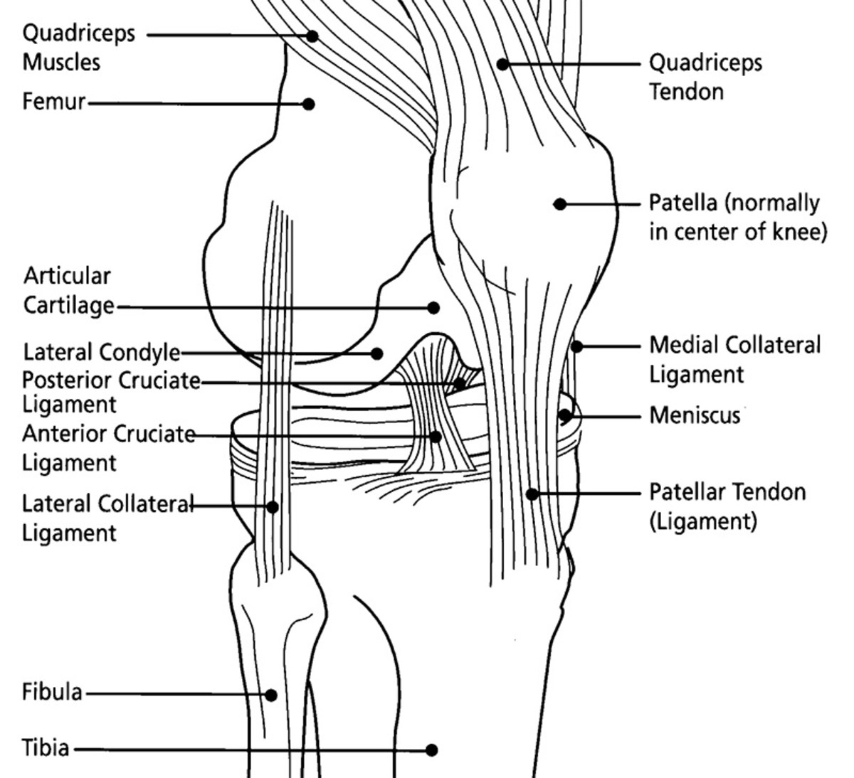
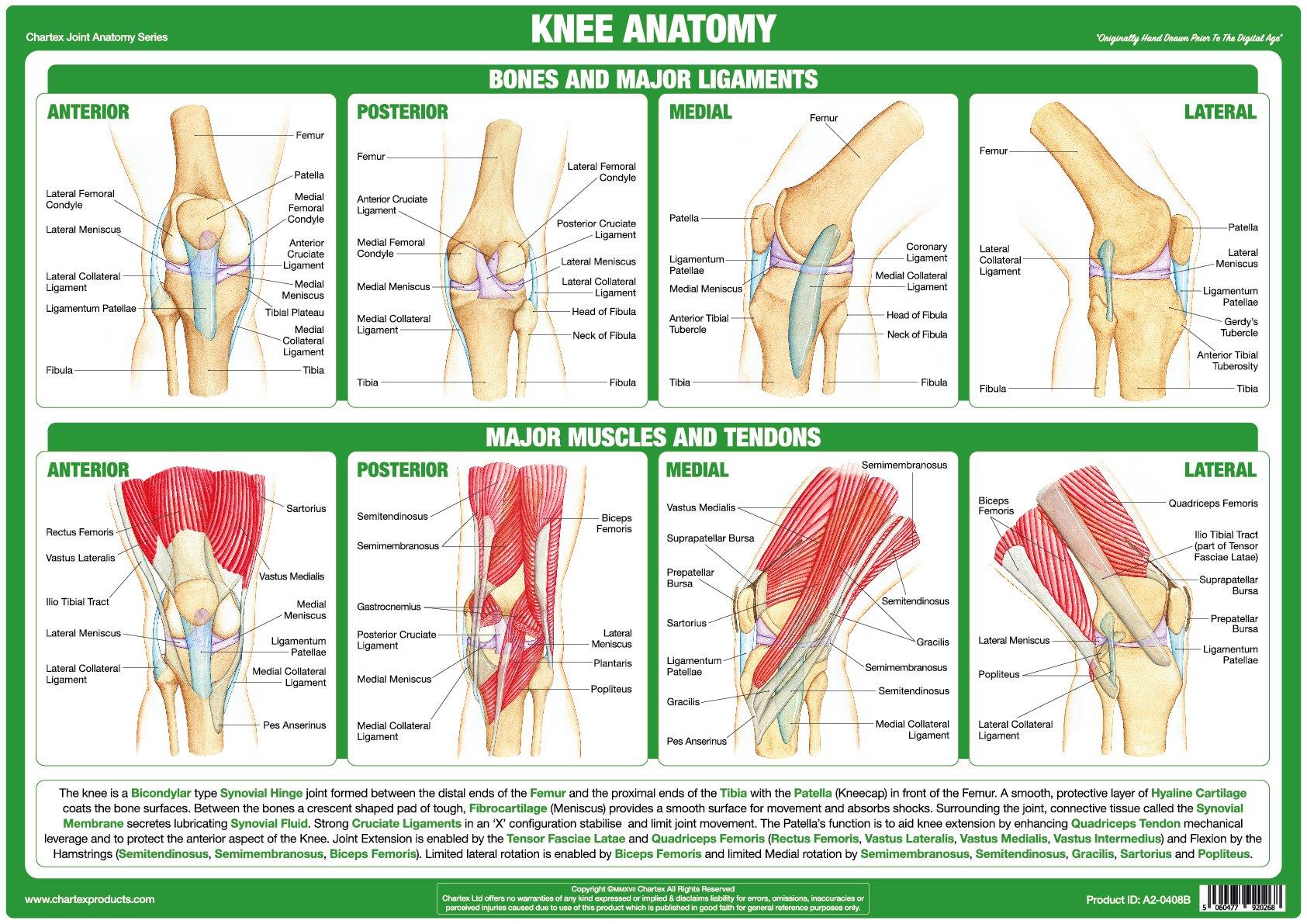
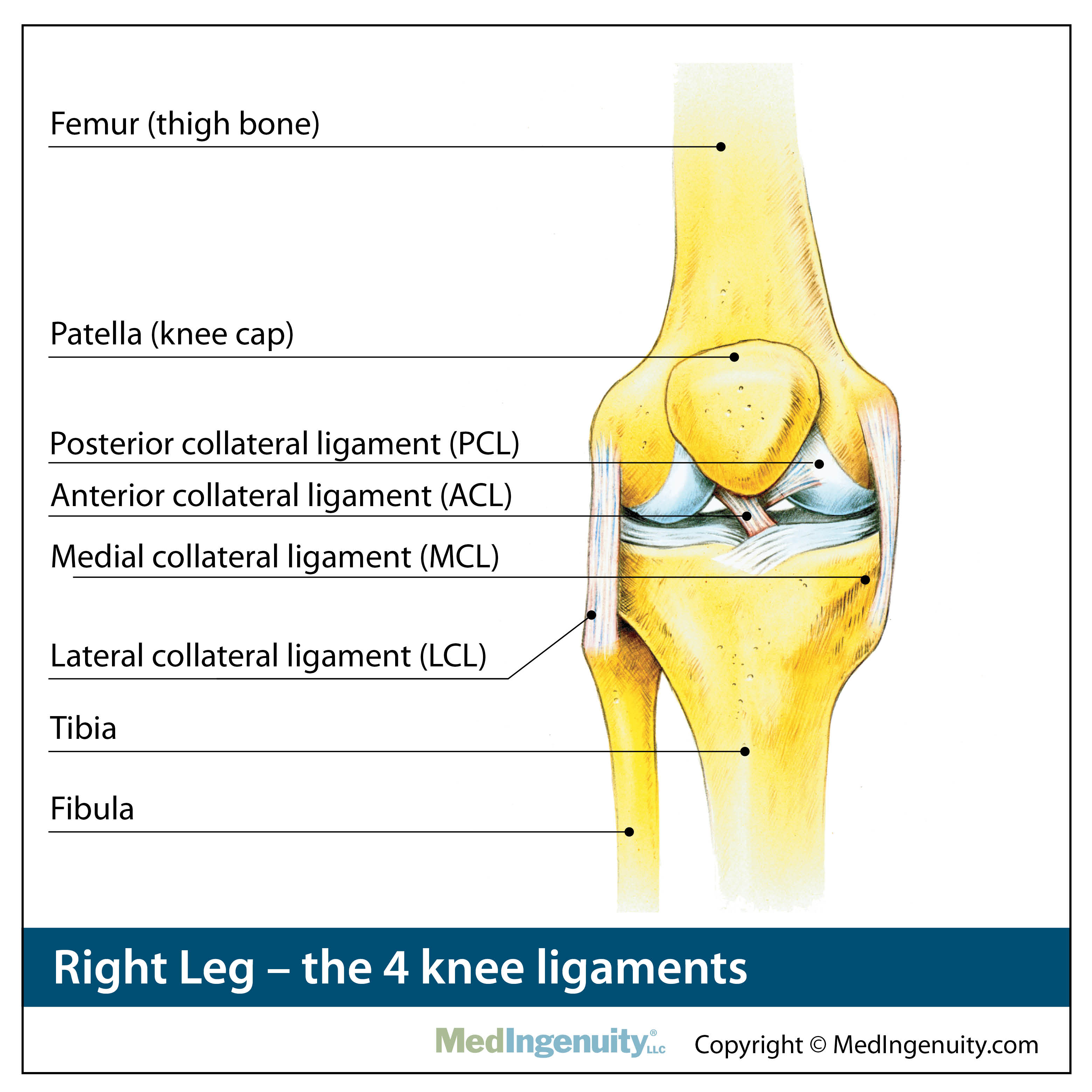
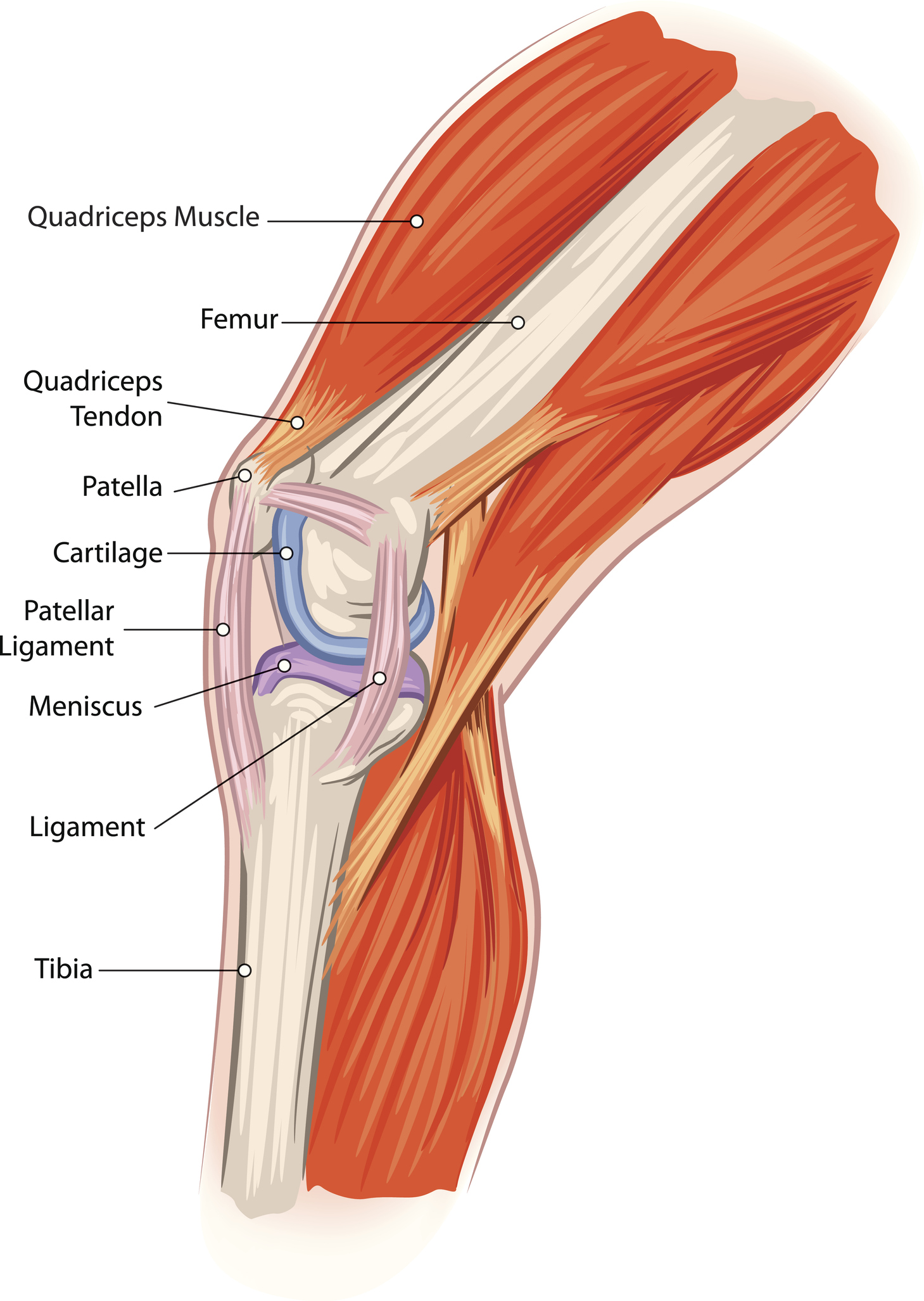
Anatomical Drawing Of The Knee - Which type of joint is the knee? This article will introduce knee joint with knee anatomy. The knee joint is a synovial joint this means it contains a fluid that lubricates it. Web the knee is the meeting place of two important bones in the leg, the femur (the thighbone) and the tibia (the shinbone). It is formed by articulations between the patella, femur and tibia. Web the knee joint is the junction of the thigh and leg. The knee joint is a synovial joint. It is a complex hinge joint composed of two articulations; Knowing about knee anatomy can help people understand how knee arthritis develops and sometimes causes pain. Web to draw the knee, begin by visualizing the bones and tendons underneath to help with the placement of landmarks. Web the knee joint is a hinge type synovial joint, which mainly allows for flexion and extension (and a small degree of medial and lateral rotation). The knee is a complex joint that flexes, extends, and twists slightly from. An epicondyle projects from each condyle. On “contrast” the user can choose the type of mri sequence: Check out these resources. The knee joint is a synovial joint. Web this article takes a concise look at the anatomy of the knee joint and describes the processes and conditions that cause pain in the different aspects (parts) of the knee. Web to understand the function and structure of the knee joint, a knee anatomy can be helpful. There are prominent lateral and. Then draw the quadriceps muscles, and indicate the patella and its tendon down to the lower leg. The knee is the joint in the middle of your leg. The bones articulating at the knee are large and complex. Web to draw the knee, begin by visualizing the bones and tendons underneath to help with the placement of landmarks. The femur,. Finally, draw in the hamstrings covering the calves at the back of the knee. They are attached to the femur (thighbone), tibia (shinbone), and fibula (calf bone) by fibrous tissues called. In the knee joint, the femur articulates with the tibia and the patella. The patella (or kneecap, as it is commonly called) is made of bone and sits in. Web anatomy of the knee joint. Check out these resources i've made to help you learn! The structure of a normal knee joint. In this page, we will take a look at all of the above as well as the anatomy of the knee. Web the knee joint is the largest joint in the body and connects the thigh with. This fluid is known as the synovial fluid. Web structure and function. External ligaments of the knee a. The patella (or kneecap, as it is commonly called) is made of bone and sits in front of the knee. The tibiofemoral joint and patellofemoral joint. Learn about the muscles, tendons, bones, and ligaments that comprise the knee joint anatomy. Web the muscles that affect the knee’s movement run along the thigh and calf. Knowing about knee anatomy can help people understand how knee arthritis develops and sometimes causes pain. Web the knee joint is a hinge type synovial joint, which mainly allows for flexion and. Meniscus (lateral and medial), cruciate ligaments, vastus (lateralis, intermedius, medialis), tibial and fibular collateral ligaments. The knee joint is a synovial joint. The femur, tibia, and patella. Web knee joint anatomy consists of muscles, ligaments, cartilage and tendons. Web structure and function. Web the knee is the meeting place of two important bones in the leg, the femur (the thighbone) and the tibia (the shinbone). The knee joint is a synovial joint. Web anatomy of the knee on a coronal slice (mri) : Web 🧑🏽🎓learning anatomy & physiology? It’s where your thigh bone (femur) meets your shin bone (tibia). Web anatomy of the knee on a coronal slice (mri) : The thigh bone ( femur ), the shin bone ( tibia) and the kneecap ( patella) articulate through tibiofemoral and patellofemoral joints. There are prominent lateral and medial condyles at the distal end of the femur. Web the knee joint is a hinge type synovial joint, which mainly allows. Then draw the quadriceps muscles, and indicate the patella and its tendon down to the lower leg. Web anatomy of the knee joint. The knee is made up of 4 bones, but it is composed primarily of 2 joints. The patella (or kneecap, as it is commonly called) is made of bone and sits in front of the knee. The knee joint is a synovial joint. Where is the knee joint located? Web the knee joint is a hinge type synovial joint, which mainly allows for flexion and extension (and a small degree of medial and lateral rotation). The bones articulating at the knee are large and complex. The femur has a slight medial slant, while the tibia is nearly vertical. It is made up of two joints, the tibiofemoral joint (between the tibia and the femur), and the patellofemoral joint (between the patella and the femur). Web to understand the function and structure of the knee joint, a knee anatomy can be helpful. They are attached to the femur (thighbone), tibia (shinbone), and fibula (calf bone) by fibrous tissues called. Web the main features of the knee anatomy include bones, cartilages, ligaments, tendons and muscles. It is a complex hinge joint composed of two articulations; The knee joint is a synovial joint this means it contains a fluid that lubricates it. The structure of a normal knee joint.
The Complete Guide to Knee Anatomy

Knee Anatomy

Anatomy of Knee

Knee injuries causes, types, symptoms, knee injuries prevention & treatment

Anatomy of the Knee Joint (With Diagrams and XRay) Owlcation

Knee injuries causes, types, symptoms, knee injuries prevention & treatment

Knee Joint Anatomy Poster

The Knee Joint Anatomy

Anatomy, Pathology & Treatment of the Knee Joint Articles & Advice
![Runner’s Knee Causes And Treatment [𝗣]𝗥𝗲𝗵𝗮𝗯](https://i1.wp.com/theprehabguys.com/wp-content/uploads/2020/08/knee-anatomy.png?ssl=1)
Runner’s Knee Causes And Treatment [𝗣]𝗥𝗲𝗵𝗮𝗯
Web Anatomy Of The Knee On A Coronal Slice (Mri) :
Web The Knee Joint Is The Largest Joint In The Body And Connects The Thigh With The Lower Leg.
There Are Prominent Lateral And Medial Condyles At The Distal End Of The Femur.
Web The Knee, Also Known As The Tibiofemoral Joint, Is A Synovial Hinge Joint Formed Between Three Bones:
Related Post: